Fraud Blocker Skip to main content

Protist Collection (page 5)

"Exploring the Intricate World of Protists: From Seaweed Specimens to Malarial Parasites" In this captivating collection, we delve into the fascinating realm of protists

Background imageProtist Collection: Dictyota atomaria, seaweed

Dictyota atomaria, seaweed

Background imageProtist Collection: Chordaria flagellifornis, seaweed

Chordaria flagellifornis, seaweed
Plate 57 from Algae Danmonienses : or dried specimens of Marine Plants, principally collected in Devonshire by Mary Wyatt. 1834-1840

Background imageProtist Collection: Asperoccus turneri, seaweed

Asperoccus turneri, seaweed
Plate 59 from Algae Danmonienses : or dried specimens of Marine Plants, principally collected in Devonshire by Mary Wyatt. 1834-1840

Background imageProtist Collection: Fucus agarum, kelp

Fucus agarum, kelp
Plate 75 from Fuci, or coloured figures and descriptions of the Plants referred by botanists to the genus Fucus (1808-1819), Volume II, by Mary Dawson Turner

Background imageProtist Collection: Chondus cripus, carrageen moss

Chondus cripus, carrageen moss
Illustration from Algae Danmonienses: or dried specimens of Marine Plants, principally collected in Devonshire by Mary Wyatt; carefully named according to Dr. Hookers British Flora

Background imageProtist Collection: Rhodophyta, Coralline algae

Rhodophyta, Coralline algae
This specimen was collected by Charles Darwin in 1836 from Keeling Atoll, Indian Ocean

Background imageProtist Collection: Raphiidonema faringdone, calcareous sponge

Raphiidonema faringdone, calcareous sponge
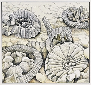
A vase-shaped calcareous sponge with numerous small canals from the Cretaceous of Berkshire, England

Background imageProtist Collection: Coccoliths magnified a thousand times

Coccoliths magnified a thousand times
An illustration of Coccoliths magnified a thousand times. Coccoliths are micro-fossils and feature heavily in the composition of chalk

Background imageProtist Collection: Lichens

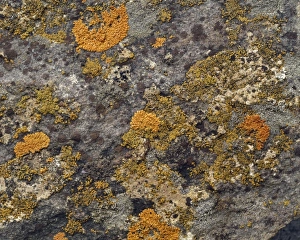
Lichens result from an intimate relationship between a fungus and an alga; there are about 18, 000 species

Background imageProtist Collection: Minakatella longifila, slime mould

Minakatella longifila, slime mould

Background imageProtist Collection: Actinoptychus, diatom

Actinoptychus, diatom
Scanning electron microscope image of the exterior valve of the diatom Actinoptychus (x 500 on a standard 9 cm wide print)

Background imageProtist Collection: Chalk

Chalk
A piece of flintless white chalk from the Upper Chalk, Flamborough, Yorks. Chalk is a sedimentary rock formed in deep seas

Background imageProtist Collection: Light Micrograph: Ciliate - Magnification x 750 (when printed A4, 29. 7 cm wide)

Light Micrograph: Ciliate - Magnification x 750 (when printed A4, 29. 7 cm wide)
LRDS-315 Light Micrograph: Ciliate Magnification x 750 (when printed A4, 29.7 cm wide) Frontonia sp. David Spears (Last Refuge) / ardea.com Last Refuge contact details: prints@ardea.com tel

Background imageProtist Collection: Pl ton -(Noctiluca scintillans) group / one with ingested copepod / x12

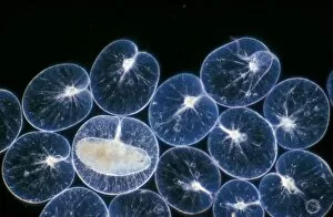
Pl ton -(Noctiluca scintillans) group / one with ingested copepod / x12

Background imageProtist Collection: Metopus protozoan

Metopus protozoan

Background imageProtist Collection: Thalassomyxa australis protozoan

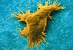
Thalassomyxa australis protozoan
Thalassomyxa protozoan. Coloured scanning electron micrograph (SEM) of a Thalassomyxa australis prot- ozoan (single-celled animal)

Background imageProtist Collection: Lembadion protozoan

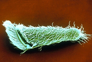
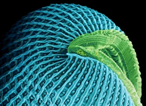
Lembadion protozoan. Coloured scanning electron micrograph (SEM) of a Lembadion bullinum protozoan (single-celled animal)

Background imageProtist Collection: Orbulina foraminiferan, SEM

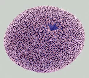
Orbulina foraminiferan, SEM
Orbulina. Coloured scanning electron micrograph (SEM) of the shell of the foraminiferan Orbulina sp. Foraminiferans are marine single-celled protists that construct and inhabit shells (tests)

Background imageProtist Collection: Euplotes ciliate protozoan, SEM

Euplotes ciliate protozoan, SEM
Euplotes ciliate protozoan. Coloured scanning electron micrograph (SEM) of a ciliate protozoan (Euplotes sp.). This single-celled organism is found in fresh and marine water

Background imageProtist Collection: Spirochona protozoa

Spirochona protozoa

Background imageProtist Collection: Ciliate protozoans, SEM

Ciliate protozoans, SEM
Ciliate protozoans. Coloured scanning electron micrograph (SEM) of sessile (anchored) ciliate protozoan (subclass Peritrichia)

Background imageProtist Collection: Trichomytopsis protozoan

Trichomytopsis protozoan

Background imageProtist Collection: Ciliate protozoa, SEM

Ciliate protozoa, SEM
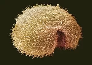
Ciliate protozoa. Coloured scanning electron micrograph (SEM) of cilliate protozoa (unicellular animals). The ciliates are so named because of the hair-like cilia surrounding their bodies which are

Background imageProtist Collection: Breslauides ciliate protozoan, SEM

Breslauides ciliate protozoan, SEM
Breslauides ciliate protozoan. Coloured scanning electron micrograph (SEM) of a ciliate protozoan (Breslauides sp.). This tiny single-celled organism is found in water

Background imageProtist Collection: Tetrahymena protozoa

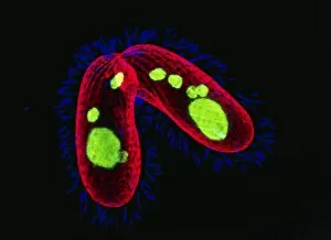
Tetrahymena protozoa. Immunofluorescent light micrograph of two Tetrahymena thermophila protozoa (single-celled animals). Nuclei are green, cell walls red and cilia (hairs) blue. T

Background imageProtist Collection: Dendrocometes protozoan

Dendrocometes protozoan. Coloured scanning electron micrograph (SEM) of a Dendrocometes paradoxus protozoan (single-celled animal)

Background imageProtist Collection: Blepharisma ciliate protozoan, SEM

Blepharisma ciliate protozoan, SEM
Blepharisma ciliate protozoan. Coloured scanning electron micrograph (SEM) of a ciliate protozoan (Blepharisma sp.). This tiny single- celled organism is found in freshwater

Background imageProtist Collection: Foraminiferans, light micrograp

Foraminiferans, light micrograp
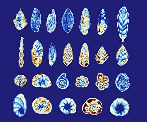
Foraminiferans, polarised light micrograph. Foraminiferans are marine single-celled protists that construct and inhabit shells (tests)

Background imageProtist Collection: Powdery scab infection, light micrograph

Powdery scab infection, light micrograph
Powdery scab infection. Light micrograph of a section through a potato infected with powdery scab (Spongospora subterranea), showing pustules and spores. S

Background imageProtist Collection: Clubroot infection, light micrograph

Clubroot infection, light micrograph
Clubroot infection. Light micrograph a section through a cabbage root infected with clubroot (Plasmodiophora brassicae). Plasmodiophora is a parasitic protist that infects the roots of cabbages

Background imageProtist Collection: Radiolarian, light micrograph

Radiolarian, light micrograph
Radiolarian, differential interference contrast micrograph. Radiolaria are single-celled protozoans that are found in marine plankton

Background imageProtist Collection: Radiolaria, light micrograph

Radiolaria, light micrograph
Radiolaria. Light micrograph of various radiolaria. Radiolaria are single-celled protozoans that are found in marine plankton

Background imageProtist Collection: Paramecium protozoan, artwork

Paramecium protozoan, artwork
Paramecium protozoan. Computer artwork of a Paramecium sp. ciliate protozoan. Its single cell is covered in numerous cilia (white lines), hair-like structures that it beats for propulsion

Background imageProtist Collection: Loxophyllum ciliate, light micrograph

Loxophyllum ciliate, light micrograph
Loxophyllum ciliate. Light micrograph of a Loxophyllum helus ciliate protozoan. This tiny single-celled organism is found in marine environments

Background imageProtist Collection: Protozoa, historical artwork

Protozoa, historical artwork
Protozoa. 1839 artwork by the naturalist Christian Gottfried Ehrenberg of Protozoa as seen under his microscope

Background imageProtist Collection: Trichodina parasite, SEM

Trichodina parasite, SEM
Trichodina parasite. Coloured scanning electron micrograph (SEM) of a Trichodina sp. protist on a freshwater minnow. This ciliate parasite lives on the skin

Background imageProtist Collection: Actinosphaerium protozoan

Actinosphaerium protozoan. Differential interference contrast micrograph of an Actinoshaerium sp. heliozoa protozoan. This single-celled freshwater organism has many pseudopodia radiating outwards

Background imageProtist Collection: Euglena protozoan, light micrograph

Euglena protozoan, light micrograph
Euglena protozoan. Differential interference contrast micrograph of the flagellate protozoan Euglena spirogyra. This freshwater single-celled organism can either obtain energy from sunlight

Background imageProtist Collection: Euglena protozoa, light micrograph

Euglena protozoa, light micrograph
Euglena protozoa. Differential interference contrast micrograph of the flagellate protozoa Euglena acus. These freshwater single-celled organisms can either obtain energy from sunlight

Background imageProtist Collection: Ciliate protozoan, SEM

Ciliate protozoan, SEM
Ciliate protozoan. Coloured scanning electron micrograph (SEM) of a Paramecium cilliate protozoan. Protozoa are a type of unicellular animal

Background imageProtist Collection: Selection of diatoms, SEM

Selection of diatoms, SEM
Selection of diatoms, coloured scanning electron micrograph (SEM). Diatoms are single-celled photosynthetic algae, of which there are about 100, 000 species

Background imageProtist Collection: Ciliate protozoan membranelles, SEM

Ciliate protozoan membranelles, SEM
Ciliate protozoan membranelles, coloured scanning electron micrograph (SEM). Protozoa are a type of unicellular animal. The ciliate protozoa are so named because of the hair-like cilia which are used

Background imageProtist Collection: Coscinodiscus diatom, SEM

Coscinodiscus diatom, SEM
Coscinodiscus diatom. Coloured scanning electron micrograph (SEM) of a Coscinodiscus diatom (red). This is a marine diatom


All Professionally Made to Order for Quick Shipping

"Exploring the Intricate World of Protists: From Seaweed Specimens to Malarial Parasites" In this captivating collection, we delve into the fascinating realm of protists. The pressed seaweed specimens C016/6127 reveal a stunning diversity, with diatoms taking center stage under the scanning electron microscope (SEM). Dictyota dichotoma showcases its intricate branching structure, reminiscent of an artistic masterpiece. Transporting us back in time is the Rye Beach, New Hampshire Postcard from 1903. This vintage gem captures the beauty of Fucus bulbosus, a majestic kelp swaying in the ocean currents. Another diatom steals our attention under SEM – Fucus radiatus displays its delicate fronds and intricate patterns. But not all they are as visually pleasing; Plasmodium sp. , a malarial parasite, reminds us of their impact on human health. Acanthophracta radiolarians mesmerize us with their intricately sculpted skeletons that resemble miniature works of art. The light micrograph (LM) reveals a hidden world within protozoans - a kidney-shaped ciliate surrounded by Euglena sp. , both magnified x900 when printed A4 size. It's astonishing how much detail can be captured at such high magnification. Calcareous alga Coelosphaeridium adds another dimension to this diverse group while diatoms continue to amaze with their varied shapes and structures. Protists truly showcase nature's creativity and complexity on microscopic scales. Join us as we unravel their secrets and appreciate these tiny wonders that play vital roles in our ecosystems.