Fraud Blocker Skip to main content

Protist Collection (page 2)

"Exploring the Intricate World of Protists: From Seaweed Specimens to Malarial Parasites" In this captivating collection, we delve into the fascinating realm of protists

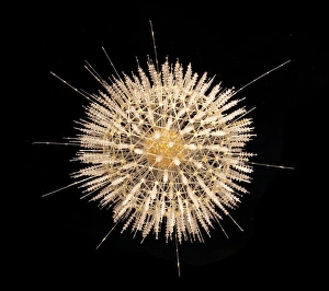
Background imageProtist Collection: Aulosphaera elegantissima, radiolarian

Aulosphaera elegantissima, radiolarian
A glass model of a radiolarian, created by Leopold and Rudolf Blaschka in the late nineteenth century and held at the Natural History Museum, London

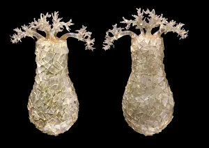
Background imageProtist Collection: Difflugia pyriformis, amoebae

Difflugia pyriformis, amoebae
A glass model of amoebae, created by Leopold and Rudolf Blaschka in the late nineteenth century and held at the Natural History Museum, London

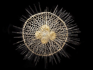
Background imageProtist Collection: Radiolarian

Radiolarian
A glass model of a radiolarian, created by Leopold and Rudolf Blaschka in the late nineteenth century and held at the Natural History Museum, London

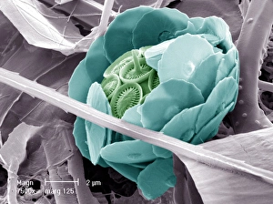
Background imageProtist Collection: Syracosphaera anthos

Syracosphaera anthos
Coccosphere from the Western Mediterranean. False coloured to show the shell is formed of inner and outer layers of coccoliths with very different structure

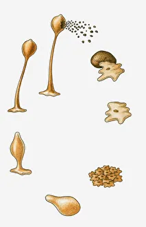
Background imageProtist Collection: Illustration of reproduction cycle of slime mould (protist)

Illustration of reproduction cycle of slime mould (protist)

Background imageProtist Collection: Picture No. 11675491

Picture No. 11675491
Light Micrograph: Ciliate. Date:

Background imageProtist Collection: Picture No. 11675490

Picture No. 11675490
Light Micrograph (LM): Elongate Ciliate and Paramecium bursaria. Date:

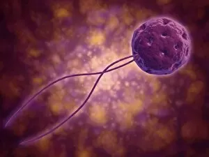
Background imageProtist Collection: Conceptual image of a viral spore

Conceptual image of a viral spore

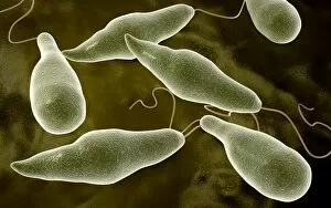
Background imageProtist Collection: Conceptual image of Euglena

Conceptual image of Euglena

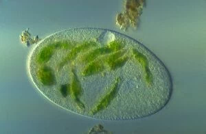
Background imageProtist Collection: Microscopic view of Paramecium bursaria

Microscopic view of Paramecium bursaria, a species of ciliate protozoan that has a mutualistic symbiotic relationship with green algae called Zoochlorella

Background imageProtist Collection: Microscopic view of protozoa

Microscopic view of protozoa. Leishmaniasis and dysentery are examples of diseases involving protozoa, often transmitted to humans by the bite of a parasite-carrying insect

Background imageProtist Collection: Giants Stairs, Baileys Island, Portland, ME. Postcard. ca. 1904-1905, Giants Stairs

Giants Stairs, Baileys Island, Portland, ME. Postcard. ca. 1904-1905, Giants Stairs, Baileys Island, Portland, ME. Postcard

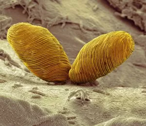
Background imageProtist Collection: Chilodonella ciliate protozoan, SEM

Chilodonella ciliate protozoan, SEM
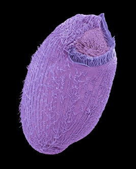
Chilodonella ciliate protozoa, coloured scanning electron micrograph (SEM). These tiny single- celled organisms are found in fresh water

Background imageProtist Collection: Holosticha ciliate protozoan, SEM

Holosticha ciliate protozoan, SEM
Holosticha ciliate protozoan. Coloured scanning electron micrograph (SEM) of a Holosticha sp. ciliate protozoan ingesting diatoms (blue). Holosticha is a tiny single-celled aquatic organism

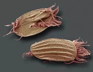
Background imageProtist Collection: Euplotes protozoa, SEM C016 / 9040

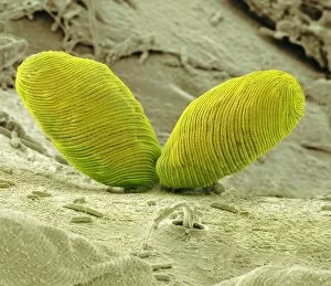
Euplotes protozoa, SEM C016 / 9040
Euplotes protozoa. Coloured scanning electron micrograph (SEM) showing the upperside (bottom) and underside (top) of two Euplotes sp. hypotrich ciliate protozoa

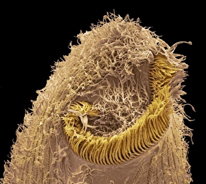
Background imageProtist Collection: Climacostomum protozoan, SEM C016 / 9063

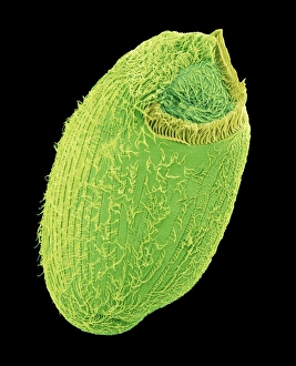
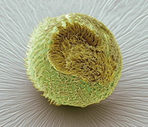
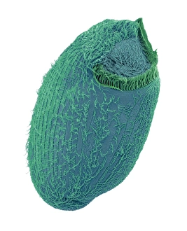
Climacostomum protozoan, SEM C016 / 9063
Climacostomum protozoan. Coloured scanning electron micrograph (SEM) of a Climacostomum sp. unicellular ciliate protozoan, showing the cells large oral apparatus (round, centre)

Background imageProtist Collection: Spirogyra algae, light micrograph C016 / 9592

Spirogyra algae, light micrograph C016 / 9592
Spirogyra algae. Polarised light micrograph of Spirogyra sp. algae. This filamentous green algae is named for the spiral arrangement of its chloroplasts (green)

Background imageProtist Collection: Spirogyra algae, light micrograph C016 / 9594

Spirogyra algae, light micrograph C016 / 9594
Spirogyra algae. Rheinberg illuminated light micrograph of Spirogyra sp. algae. This filamentous green algae is named for the spiral arrangement of its chloroplasts (green)

Background imageProtist Collection: Euglena flagellate protozoa, SEM C016 / 9103

Euglena flagellate protozoa, SEM C016 / 9103
Euglena flagellate protozoa. Coloured scanning electron micrograph (SEM) of two Euglena sp. flagellate protozoa. Euglena sp

Background imageProtist Collection: Desmids and spirogyra, light micrograph C016 / 9595

Desmids and spirogyra, light micrograph C016 / 9595
Desmids and spirogyra. Polarised light micrograph of two Micrasterias rotata desmids (round) and a single filament of Spirogyra sp. green alga (lower right)

Background imageProtist Collection: Climacostomum protozoan, SEM C016 / 9121

Climacostomum protozoan, SEM C016 / 9121
Climacostomum protozoan. Coloured scanning electron micrograph (SEM) of a Climacostomum sp. unicellular ciliate protozoan, showing the cells large oral apparatus (round, centre)

Background imageProtist Collection: Climacostomum protozoan, SEM C016 / 9061

Climacostomum protozoan, SEM C016 / 9061
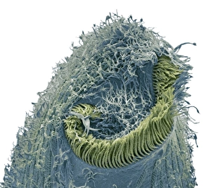
Climacostomum protozoan. Coloured scanning electron micrograph (SEM) of a Climacostomum sp. unicellular ciliate protozoan, showing the cells large oral apparatus (round, upper right)

Background imageProtist Collection: Climacostomum protozoan, SEM C016 / 9120

Climacostomum protozoan, SEM C016 / 9120
Climacostomum protozoan. Coloured scanning electron micrograph (SEM) of a Climacostomum sp. unicellular ciliate protozoan, showing the cells large oral apparatus (round, centre)

Background imageProtist Collection: Climacostomum protozoan, SEM C016 / 9065

Climacostomum protozoan, SEM C016 / 9065
Climacostomum protozoan. Coloured scanning electron micrograph (SEM) of a Climacostomum sp. unicellular ciliate protozoan, showing the cells large oral apparatus (round, upper right)

Background imageProtist Collection: Euglena flagellate protozoa, SEM C016 / 9104

Euglena flagellate protozoa, SEM C016 / 9104
Euglena flagellate protozoa. Coloured scanning electron micrograph (SEM) of two Euglena sp. flagellate protozoa. Euglena sp

Background imageProtist Collection: Climacostomum protozoan, SEM C016 / 9064

Climacostomum protozoan, SEM C016 / 9064
Climacostomum protozoan. Coloured scanning electron micrograph (SEM) of a Climacostomum sp. unicellular ciliate protozoan, showing the cells large oral apparatus (round, centre)

Background imageProtist Collection: Climacostomum protozoan, SEM C016 / 9122

Climacostomum protozoan, SEM C016 / 9122
Climacostomum protozoan. Coloured scanning electron micrograph (SEM) of a Climacostomum sp. unicellular ciliate protozoan, showing the cells large oral apparatus (round, centre)

Background imageProtist Collection: Spirogyra algae, light micrograph C016 / 9593

Spirogyra algae, light micrograph C016 / 9593
Spirogyra algae. Polarised light micrograph of Spirogyra sp. algae. This filamentous green algae is named for the spiral arrangement of its chloroplasts (green)

Background imageProtist Collection: Heliozoan, SEM C016 / 9024

Heliozoan, SEM C016 / 9024
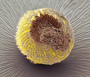
Heliozoan. Coloured scanning electron micrograph (SEM) of a heliozoan freshwater protozoan. This single-celled organism has many projections, known as axopods, radiating from its cell body

Background imageProtist Collection: Euplotes protozoa, SEM C016 / 9041

Euplotes protozoa, SEM C016 / 9041
Euplotes protozoa. Coloured scanning electron micrograph (SEM) showing the upperside (bottom) and underside (top) of two Euplotes sp. hypotrich ciliate protozoa

Background imageProtist Collection: Climacostomum protozoan, SEM C016 / 9062

Climacostomum protozoan, SEM C016 / 9062
Climacostomum protozoan. Coloured scanning electron micrograph (SEM) of a Climacostomum sp. unicellular ciliate protozoan, showing the cells large oral apparatus (round, upper right)

Background imageProtist Collection: Heliozoan, SEM C016 / 9023

Heliozoan, SEM C016 / 9023
Heliozoan. Coloured scanning electron micrograph (SEM) of a heliozoan freshwater protozoan. This single-celled organism has many projections, known as axopods, radiating from its cell body

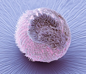
Background imageProtist Collection: Tetrahymena protozoan, SEM

Tetrahymena protozoan, SEM
Tetrahymena protozoan. Coloured scanning electron micrograph (SEM) of a Tetrahymena sp. ciliate protozoan. Tetrahymena are free-living freshwater unicellular organisms found in ponds

Background imageProtist Collection: Sleeping sickness parasites, SEM

Sleeping sickness parasites, SEM
Sleeping sickness parasites. Coloured scanning electron micrograph (SEM) of a Trypanosoma brucei parasites (blue) and white blood cells (pink) in a mouse liver. T

Background imageProtist Collection: Sleeping sickness parasite, SEM

Sleeping sickness parasite, SEM
Sleeping sickness parasite. Coloured scanning electron micrograph (SEM) of a Trypanosoma brucei parasite (blue) and a white blood cell (green) in a mouse liver. T

Background imageProtist Collection: Fossil foraminifera necklace C016 / 5995

Fossil foraminifera necklace C016 / 5995
Fossil foraminifera (Alveolina sp.) necklace from Sindh, India. The fossils are from Eocene limestone. Necklace from John Whittakers cabinet. Natural History Museum, London, UK

Background imageProtist Collection: Fungus (Lycoperdon coliforme) C016 / 5988

Fungus (Lycoperdon coliforme) C016 / 5988
Fungus (Lycoperdon coliforme). Plate 1 from Drawings of Submerged Algae (1800) by Mary Dawson Turner. Held in the Botany Library at the Natural History Museum, London, UK

Background imageProtist Collection: Dried seaweed specimens C016 / 5977

Dried seaweed specimens C016 / 5977
Dried seaweed (Chorda filum) specimens. Herbarium sheet from the collection held at the Natural History Museum, London, UK

Background imageProtist Collection: Didinium ciliate protozoan, SEM C019 / 0235

Didinium ciliate protozoan, SEM C019 / 0235
Didinium sp. ciliate protozoan, coloured scanning electron micrograph (SEM). This tiny single-celled organism is found in freshwater and marine habitats

Background imageProtist Collection: Spirostomum ciliate protozoan, SEM C019 / 0308

Spirostomum ciliate protozoan, SEM C019 / 0308
Spirostomum sp. ciliate protozoan, coloured scanning electron micrograph (SEM). These tiny single-celled organisms are found in freshwater and marine habitats

Background imageProtist Collection: Blepharisma ciliate protozoan, SEM C019 / 0228

Blepharisma ciliate protozoan, SEM C019 / 0228
Blepharisma ciliate protozoan, coloured scanning electron micrograph (SEM). These tiny single-celled organisms are found in freshwater and marine habitats

Background imageProtist Collection: Didinium ciliate protozoan, SEM C019 / 0237

Didinium ciliate protozoan, SEM C019 / 0237
Didinium sp. ciliate protozoan, coloured scanning electron micrograph (SEM). This tiny single-celled organism is found in freshwater and marine habitats

Background imageProtist Collection: Spirostomum ciliate protozoan, SEM C019 / 0307

Spirostomum ciliate protozoan, SEM C019 / 0307
Spirostomum sp. ciliate protozoan, coloured scanning electron micrograph (SEM). These tiny single-celled organisms are found in freshwater and marine habitats

Background imageProtist Collection: Seaweed (Desmarestia ligulata) C016 / 6014

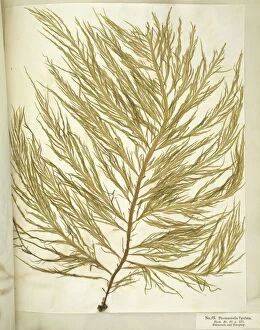
Seaweed (Desmarestia ligulata) C016 / 6014
seaweed (Desmarestia ligulata). Page 55 from Algae Danmonienses: or dried specimens of Marine Plants, principally collected in Devonshire by Mary Wyatt; carefully named according to Dr


All Professionally Made to Order for Quick Shipping

"Exploring the Intricate World of Protists: From Seaweed Specimens to Malarial Parasites" In this captivating collection, we delve into the fascinating realm of protists. The pressed seaweed specimens C016/6127 reveal a stunning diversity, with diatoms taking center stage under the scanning electron microscope (SEM). Dictyota dichotoma showcases its intricate branching structure, reminiscent of an artistic masterpiece. Transporting us back in time is the Rye Beach, New Hampshire Postcard from 1903. This vintage gem captures the beauty of Fucus bulbosus, a majestic kelp swaying in the ocean currents. Another diatom steals our attention under SEM – Fucus radiatus displays its delicate fronds and intricate patterns. But not all they are as visually pleasing; Plasmodium sp. , a malarial parasite, reminds us of their impact on human health. Acanthophracta radiolarians mesmerize us with their intricately sculpted skeletons that resemble miniature works of art. The light micrograph (LM) reveals a hidden world within protozoans - a kidney-shaped ciliate surrounded by Euglena sp. , both magnified x900 when printed A4 size. It's astonishing how much detail can be captured at such high magnification. Calcareous alga Coelosphaeridium adds another dimension to this diverse group while diatoms continue to amaze with their varied shapes and structures. Protists truly showcase nature's creativity and complexity on microscopic scales. Join us as we unravel their secrets and appreciate these tiny wonders that play vital roles in our ecosystems.