Fraud Blocker Skip to main content

Physiological Collection (page 9)

"Exploring the Intricacies of Physiology: From Airpumps to Blood Cells" Delving into the world of physiology

Background imagePhysiological Collection: Electronic and biologic systems, artwork

Electronic and biologic systems, artwork
Electronic and biological systems. Computer artwork showing a comparison between electronic and biological systems, showing similarities between the components at different levels of complexity

Background imagePhysiological Collection: Space research

Space research. Computer artwork of two satellites in Earth orbit for biological research. A mouse is seen in one of the satellites

Background imagePhysiological Collection: Swing trainer for cosmonauts

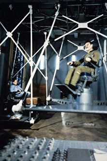
Swing trainer for cosmonauts
Swing trainer at the Gagarin Cosmonauts Training Centre in Russia. The chair spins to disorient the trainee cosmonaut in order to assess his ability to work in difficult environments

Background imagePhysiological Collection: Centrifuge training for cosmonauts

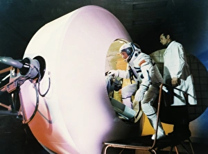
Centrifuge training for cosmonauts
Centrifuge training at the Gagarin Cosmonauts Training Centre in Russia. The centrifuge simulates the forces experienced due to acceleration at launch so that the cosmonaut can be assessed for his

Background imagePhysiological Collection: Dislocated elbow, X-ray

Dislocated elbow, X-ray
Dislocated elbow. Coloured lateral X-ray of an injury to the elbow (centre) showing that the upper arm bone (humerus, top right) is misaligned (dislocated, turquoise)

Background imagePhysiological Collection: Wound scab, SEM

Wound scab, SEM
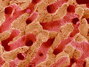
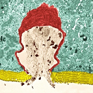
Wound scab. Coloured scanning electron micrograph (SEM) of keratinocytes (brown) lining a skin wound. Keratinocytes are skin cells that contain the protein keratin

Background imagePhysiological Collection: Fertilisation

Fertilisation, computer artwork. Sperm fertilising an egg

Background imagePhysiological Collection: Fertility

Fertility. Conceptual computer artwork of human sperm (male sex cells, white) as part of a board game. The dice and chance cards (left)

Background imagePhysiological Collection: Kidney tubule cell, coloured SEM

Kidney tubule cell, coloured SEM
Kidney tubule cell, coloured scanning electron micrograph (SEM). This cell (brown) is covered in thin projections (microvilli) that serve to increase its surface area

Background imagePhysiological Collection: Kidney tubules

Kidney tubules. Coloured scanning electron micrograph (SEM) of proximal convoluted tubules (dark brown) in the kidney. The proximal convoluted tubules function to reabsorb water

Background imagePhysiological Collection: Liver blood supply, SEM

Liver blood supply, SEM
Liver blood supply. Coloured scanning electron micrograph (SEM) of several capillaries running through liver tissue. The functions of the liver include destroying old red blood cells

Background imagePhysiological Collection: Liver cells, TEM

Liver cells, TEM
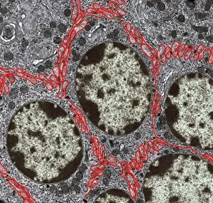
Liver cells. Coloured transmission electron micrograph (TEM) of cells in a liver, showing their tessellating boundaries (red) and nuclei (beige). Magnification: x9000 when printed 10 centimetres wide

Background imagePhysiological Collection: Goblet cell

Goblet cell. Coloured transmission electron micrograph (TEM) of a section through a goblet cell in the lining of the small intestine, part of the digestive tract

Background imagePhysiological Collection: Human ear

Human ear, computer artwork

Background imagePhysiological Collection: Vision and the brain, MRI scan

Vision and the brain, MRI scan
Vision and the brain. Coloured Magnetic Resonance Imaging (MRI) scan of an axial section through the head of a 55 year old man, showing the eyes, optic nerves and brain

Background imagePhysiological Collection: Diabetes mellitus, computer artwork

Diabetes mellitus, computer artwork. Diabetes is caused by low levels of the hormone insulin and resistance to insulin by the body cells

Background imagePhysiological Collection: The iris of the eye

The iris of the eye
Iris. Computer-enhanced image of the blue/brown iris of a human eye. The iris is a ring of muscle fibres which contracts and relaxes to alter the light entering the eye through the pupil (black)

Background imagePhysiological Collection: Iris

Iris. Computer-enhanced and coloured image of a human iris. The iris is a ring of muscle fibres. It contracts and relaxes to alter the amount of light entering the eye through the pupil (black)

Background imagePhysiological Collection: Synapse, computer artwork

Synapse, computer artwork
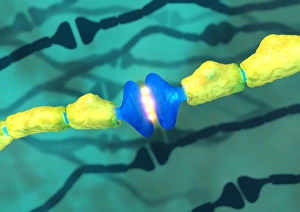
Synapses. Computer artwork of synapses, the junctions between the ends (blue, swollen) of two nerve cells (neurons). Nerve cells are responsible for passing information around the central nervous

Background imagePhysiological Collection: Nerve support cell, SEM

Nerve support cell, SEM
Nerve support cell. Coloured scanning electron micrograph (SEM) of an oligodendrocyte cell. This cell forms the myelin sheaths around nerve fibres in the central nervous system (brain and spinal cord)

Background imagePhysiological Collection: Brain and blood vessels, 3-D CT scan

Brain and blood vessels, 3-D CT scan
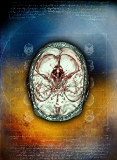
Brain and blood vessels, coloured 3-D computed tomography (CT) scan. The front of the brain is at bottom. It is enclosed by the skull (grey/green). Blood vessels (red) supply the brain with blood

Background imagePhysiological Collection: Head, side view, MRI scan

Head, side view, MRI scan
Head from the side, magnetic resonance imaging (MRI) scan. The brain (light blue, folded) fills the cerebrum of the skull (white). At centre left is an eyeball (dark blue circle)

Background imagePhysiological Collection: Heart, chest X-ray

Heart, chest X-ray

Background imagePhysiological Collection: Contour map of the heart

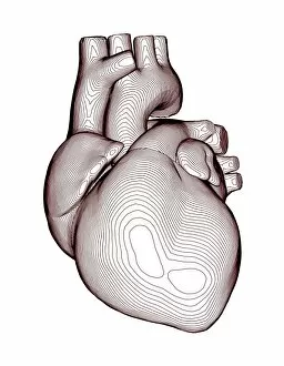
Contour map of the heart, computer artwork. The contours on this map describe the surface shape of the heart, shown here from the front

Background imagePhysiological Collection: Brain

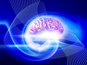
Brain. Conceptual computer artwork of a side view of a human brain emitting rays of light. The front of the brain is at right

Background imagePhysiological Collection: Macrophage cell, TEM

Macrophage cell, TEM
Macrophage. Coloured transmission electron micrograph (TEM) of a macrophage cell. The cells nucleus is bright green. Mitochondria (green) in the cells cytoplasm produce energy for the cell

Background imagePhysiological Collection: Blood clot crystals, SEM

Blood clot crystals, SEM
Blood clot crystals. Coloured scanning electron micrograph (SEM) of crystals of albumin from a blood clot. Albumin is the most abundant protein in the blood plasma

Background imagePhysiological Collection: Sketch of 3 antibodies

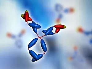
Sketch of 3 antibodies, computer artwork. An antibody, also known as an immunoglobulin, is an assembly of proteins that allows the immune system to identify

Background imagePhysiological Collection: Antibodies

Antibodies
Antibody, computer artwork. An antibody, also known as an immunoglobulin, is an assembly of proteins that allows the immune system to identify and neutralise foreign objects in the body

Background imagePhysiological Collection: Red blood cells embedded in tissue

Red blood cells embedded in tissue
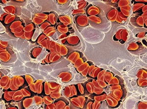
Red blood cells. Coloured scanning electron micrograph of red blood cells in tissue. Blood is composed mainly of red blood cells (erythrocytes). Red blood cells are pumped to all parts of the body

Background imagePhysiological Collection: Red blood cell

Red blood cell. Computer artwork of a human red blood cell (erythrocyte). Red blood cells are biconcave, giving them a large surface area for gas exchange, and highly elastic


All Professionally Made to Order for Quick Shipping

"Exploring the Intricacies of Physiology: From Airpumps to Blood Cells" Delving into the world of physiology, we uncover a fascinating array of scientific discoveries and visual wonders. Joseph Wright's masterpiece, "The Airpump, " captures the essence of early physiological experiments, showcasing humanity's thirst for knowledge. Intricate synapse nerve junctions depicted through Transmission Electron Microscopy (TEM) reveal the intricate communication network within our bodies. These microscopic connections enable seamless transmission of signals between neurons, shaping our thoughts and actions. Moving upwards to the head, we encounter an astonishing view of the muscular system. The complexity and precision required for even simple movements become apparent as ligaments intertwine with bones in perfect harmony. Venturing deeper into female physiology, a Scanning Electron Microscope (SEM) reveals the uterus lining during menstruation. This glimpse into nature's cyclical process sheds light on one aspect of womanhood that has both fascinated and perplexed scientists throughout history. Stepping away from biology but still within the realm of physiology, lie detector tests emerge as tools to decipher truth from deception. By monitoring physiological responses such as heart rate and perspiration levels, these tests attempt to unravel hidden truths buried within our bodies. Returning to cellular structures through TEM imaging once again unveils rough endoplasmic reticulum - a vital component responsible for protein synthesis in cells. Its intricate web-like structure showcases nature's elegance at work. Tracing back centuries ago brings us to Descartes' optics theory from the 17th century - an exploration into how vision is perceived by our eyes and interpreted by our brains. This groundbreaking theory laid foundations for understanding human perception today. Transitioning towards physicality itself leads us to skeletons intertwined with ligaments - providing structural support while allowing flexibility in movement. These silent heroes ensure smooth locomotion throughout life's journey.