Fraud Blocker Skip to main content

Microscope Collection (page 9)

"Unveiling the Unseen: Exploring the Microscopic World" Step into a realm of hidden wonders

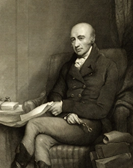
Background imageMicroscope Collection: William Hyde Wollaston, British chemist

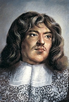
William Hyde Wollaston, British chemist
William Hyde Wollaston (1766-1828), British chemist and physicist. Wollaston gave up his medical practice in 1800 and earned a living from the sale of platinum

Background imageMicroscope Collection: Clay, SEM C015 / 5767

Clay, SEM C015 / 5767
Clay. Scanning electron microscope of a sample of clay. Clay particles are the smallest particle of the different soil types, giving the soil a smooth feel when rubbed between the fingers

Background imageMicroscope Collection: Golgi apparatus, TEM C017 / 8234

Golgi apparatus, TEM C017 / 8234
Golgi apparatus. Transmission electron micrograph (TEM) of Golgi apparatus from a Chlamydomonas sp. alga. Golgi are membrane-bound organelles that modify and package proteins

Background imageMicroscope Collection: Skeletal muscle, TEM C016 / 5369

Skeletal muscle, TEM C016 / 5369
Skeletal muscle. Transmission electron micrograph (TEM) of a cross section through human skeletal (striated) muscle. Blocks of muscle (lighter grey) are surrounded by connective tissue (black)

Background imageMicroscope Collection: FE scanning electron microscopy C016 / 3821

FE scanning electron microscopy C016 / 3821
FE scanning electron microscopy. Researcher operating a field-emission scanning electron microscope (FE-SEM). This is a F JEOL 6320F FE-SEM

Background imageMicroscope Collection: Scanning transmission electron microscopy C016 / 3815

Scanning transmission electron microscopy C016 / 3815
Scanning transmission electron microscopy. Researcher using a scanning transmission electron microscope (STEM) to analyse the structure of solid-state materials

Background imageMicroscope Collection: Tyrophagus casei, cheese mite

Tyrophagus casei, cheese mite
Scanning electron microscope image of a cheese mite (x 170). These creatures are generally considered to be a pest, however they are added to Altenburger cheese to give it flavour

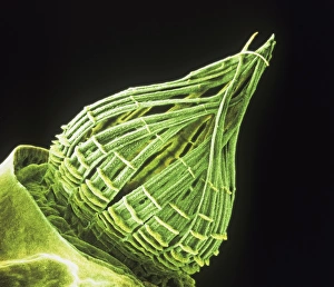
Background imageMicroscope Collection: Ceratodon purpureus, ceratodon moss spore capsule

Ceratodon purpureus, ceratodon moss spore capsule
Scanning electron microscope (SEM) image of a ceratodon moss spore capsule (x 650 on a standard 9 cm wide print)

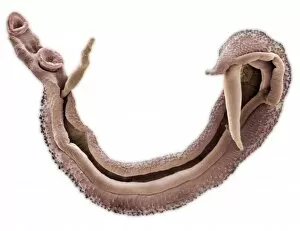
Background imageMicroscope Collection: Schistosoma nasale, bloodfluke

Schistosoma nasale, bloodfluke
Scanning electron microscope image of a parasitic bloodfluke or flatworm. Coloured artifically by computer

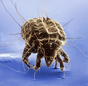
Background imageMicroscope Collection: Dermatophagoides pteronyssius, dust mite

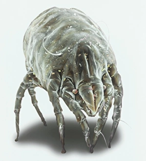
Dermatophagoides pteronyssius, dust mite
Scanning electron microscope image showing a dust mite (x 250 on standard 9cm wide print). This image has been artificially coloured by a computer

Background imageMicroscope Collection: Euglena gracilis

Euglena gracilis protists. Light micrograph of a group of Euglena gracilis protists. These organisms possess a unique combination of plant and animal characters. Like the algae, E

Background imageMicroscope Collection: Lung alveoli

Lung alveoli. Light micrograph of a section through normal human lung tissue showing alveoli. These are tiny air sacs where most of the diffusion of gases to and from the blood occurs

Background imageMicroscope Collection: Cardiac muscle, TEM

Cardiac muscle, TEM
Cardiac muscle. Coloured transmission electron micrograph (TEM) of cardiac muscle fibrils (orange)from a healthy heart. Mitochondria (pink) supply the muscle cells with energy

Background imageMicroscope Collection: Cardiac muscle and capillary, TEM

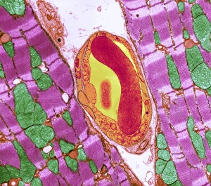
Cardiac muscle and capillary, TEM
Cardiac muscle. Coloured transmission electron micrograph (TEM) of cardiac muscle fibrils (purple)from a healthy heart. Mitochondria (green) supply the muscle cells with energy

Background imageMicroscope Collection: Cell biology laboratory

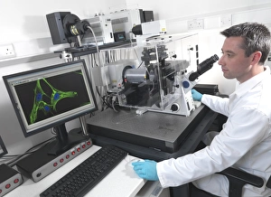
Cell biology laboratory. Scientist using confocal fluorescence microscopy to view stem cells

Background imageMicroscope Collection: Smallpox virus particle, TEM

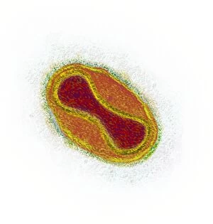
Smallpox virus particle, TEM
Smallpox virus particle. Coloured transmission electron micrograph (TEM) of a Variola major virus particle. Each particle consists of a DNA (deoxyribonucleic acid)

Background imageMicroscope Collection: Infectious bronchitis virus (IBV), TEM

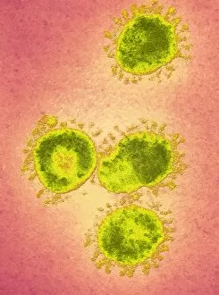
Infectious bronchitis virus (IBV), TEM
Infectious bronchitis virus (IBV) particles, coloured transmission electron micrograph (TEM). This virus is a member of the coronavirus family

Background imageMicroscope Collection: Eastern equine encephalitis virus, TEM

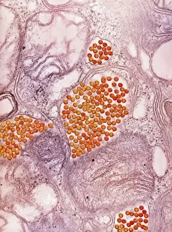
Eastern equine encephalitis virus, TEM
Eastern equine encephalitis virus. Coloured transmission electron micrograph (TEM) of eastern equine encephalitis (EEE) virus particles (Alphavirus sp. orange) in the salivary gland of a mosquito

Background imageMicroscope Collection: HIV particles, TEM

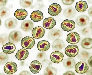
HIV particles, TEM
HIV (human immunodeficiency virus) particles, coloured transmission electron micrograph (TEM). Each particle consists of an RNA (ribonucleic acid, purple/brown) genome

Background imageMicroscope Collection: Boy Using Microscope

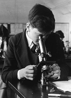
Boy Using Microscope
A schoolboy at Lingfield Secondary uses a microscope

Background imageMicroscope Collection: ROBERT HOOKE 1635 - 1703

ROBERT HOOKE 1635 - 1703
ROBERT HOOKE English scientist. Author of Micrographia (1665), in which he published results of his microscopic investigations

Background imageMicroscope Collection: Robert Hooke / Microscope

Robert Hooke / Microscope
Robert Hookes microscope

Background imageMicroscope Collection: Microscopes and microscopical objects, 1750. I: Wilsons pocket microscope. II

Microscopes and microscopical objects, 1750. I: Wilsons pocket microscope. II: Scroll microscope. III: Tripod microscope - improved form of Marshalls double microscope

Background imageMicroscope Collection: Oscillatoria, light microscope view of cyanobacteria

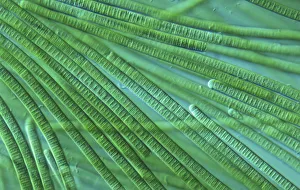
Oscillatoria, light microscope view of cyanobacteria

Background imageMicroscope Collection: A colony of bacteria seen under a microscope

A colony of bacteria seen under a microscope

Background imageMicroscope Collection: Adams Universal Microscope

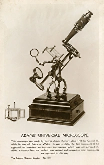
Adams Universal Microscope - made by George Adams Snr. about 1755 for King George III, while he was still Prince of Wales

Background imageMicroscope Collection: Binocular microscope large Best. London, around 1890

Binocular microscope large Best. London, around 1890
Binocular microscope large Best. Signed: R. & J. Beck Ltd London 19901. London, around 1890. The Large Best microscope was the top product of R. and J

Background imageMicroscope Collection: Transmission electron microscope EM9. Signed: Carl Zeiss

Transmission electron microscope EM9. Signed: Carl Zeiss. 1964

Background imageMicroscope Collection: Alexander Dickson

Alexander Dickson
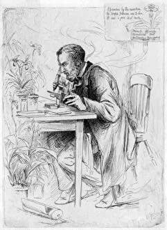
ALEXANDER DICKSON botanist, at work with his microscope : A primrose by the rivers brim, no simple primrose was to him, it was a good deal more. Date: 1836 - 1887

Background imageMicroscope Collection: Scientific Conversazione

Scientific Conversazione
The Society of Apothecaries holds a Scientific conversazione at their council chamber, Apothecaries Hall, in Blackfriars, London. The evening focused on the use of the microscope. Date: 11 April 1855

Background imageMicroscope Collection: Jean Rostand / Photo

Jean Rostand / Photo
JEAN ROSTAND French biologist and writer Date: 1894 - 1977

Background imageMicroscope Collection: Lumiere / Louis / Ils Photo

Lumiere / Louis / Ils Photo
LOUIS-JEAN LUMIERE French chemist, industrialist and pioneer (with his brother) of cinematography Date: 1864 - 1948

Background imageMicroscope Collection: Water flea, Cyclops cyprinaceus

Water flea, Cyclops cyprinaceus.. Illustration drawn and engraved by Richard Polydore Nodder. Handcolored copperplate engraving from George Shaw and Frederick Nodders The Naturalists Miscellany

Background imageMicroscope Collection: Threads of Dutch linen and gold under a microscope

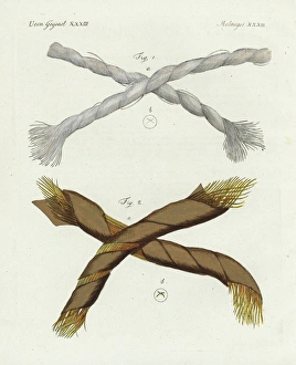
Threads of Dutch linen and gold under a microscope.. Threads of Dutch linen 1 and gold 2 magnified under a microscope. Handcolored copperplate engraving from Friedrich Bertuchs Bilderbuch fur Kinder

Background imageMicroscope Collection: Crystals of silver solution and Dianas tree

Crystals of silver solution and Dianas tree, camphor, and niter.. Crystals magnified under a microscope- silver solution and Dianas tree 1, camphor 2, and niter 3

Background imageMicroscope Collection: Human finger, skin, blood, serum and salt crystals

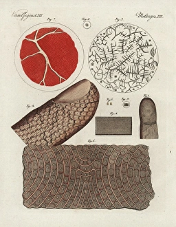
Human finger, skin, blood, serum and salt crystals.. Human finger 1, magnified 2, epidermis 3, magnified 4, scales 5, magnified skin 6, and human blood 7, serum 8

Background imageMicroscope Collection: Spermatazoa of rabbit, dog, goat and rooster

Spermatazoa of rabbit, dog, goat and rooster.. Handcolored engraving on steel by Annedouche after a drawing by Edouard Travies from Richards New Edition of the Complete Works of Comte de Buffon

Background imageMicroscope Collection: Crystals of verdigris, boric acid, common salt

Crystals of verdigris, boric acid, common salt, and sal ammoniac.. Crystals of verdigris 1, boric acid 2, common salt 3, and sal ammoniac 4 under the microscope

Background imageMicroscope Collection: LEEUWENHOEK, Antony van (1632-1723). Dutch naturalist

LEEUWENHOEK, Antony van (1632-1723). Dutch naturalist and optician. First to observe bacteriae. He is commonly known as the Father of Microbiology, and considered to be the first microbiologist. Oil

Background imageMicroscope Collection: Suffragette Flora Drummond Stanhope Toy

Suffragette Flora Drummond Stanhope Toy
A Stanhope or optical toy, a device which allows the viewing of microphotographs without using a microscope. The stanhope was invented in 1857 by Rene Dagron, a French photographer

Background imageMicroscope Collection: Boy with microscope

Boy with microscope
A boy in white overalls, sitting at a table examining something through a microscope. Date: 20th century

Background imageMicroscope Collection: Plague scientist in China

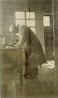
Plague scientist in China
Western doctor or scientist in China during the bubonic plague outbreak; died during research work. Date: 1911

Background imageMicroscope Collection: Boys with Microscope

Boys with Microscope
Two schoolboys with a microscope in a classroom laboratory. Date: early 1970s

Background imageMicroscope Collection: Coffee under microscope

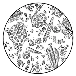
Coffee under microscope
Illustration of a coffee under microscope

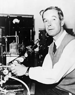
Background imageMicroscope Collection: ALAN HODGKIN (1914-1998). English physiologist. Seated at microscope. Photograph, 1963

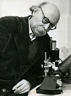
ALAN HODGKIN (1914-1998). English physiologist. Seated at microscope. Photograph, 1963

Background imageMicroscope Collection: JANE COOKE WRIGHT (1919-2013). American surgeon and cancer researcher. Photograph by Irwin Gooen

JANE COOKE WRIGHT (1919-2013). American surgeon and cancer researcher. Photograph by Irwin Gooen, 1958

Background imageMicroscope Collection: HOWARD UNIVERSITY, c1900. A class in the bacteriology laboratory at Howard University

HOWARD UNIVERSITY, c1900. A class in the bacteriology laboratory at Howard University in Washington, D.C. Photograph, c1900

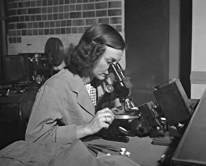
Background imageMicroscope Collection: MICROSCOPE, c1943. A woman using a microscope. Photograph, c1943

MICROSCOPE, c1943. A woman using a microscope. Photograph, c1943


All Professionally Made to Order for Quick Shipping

"Unveiling the Unseen: Exploring the Microscopic World" Step into a realm of hidden wonders, where scientists like Rosalind Franklin and Santiago Ramon y Cajal have unraveled the mysteries through their microscopic lens. From Liver Fluke to Norovirus particles, these pioneers have paved the way for groundbreaking discoveries. Witness the intricate beauty of Snail teeth and Fat cells as they come alive under the microscope's gaze. Marvel at Lubbock's Graphic 84, a visual masterpiece capturing the essence of this microcosmic universe. Delve deeper into this microscopic world and encounter Plasmodium sp. , a malarial parasite that has plagued humanity for centuries. Explore E. Coli bacterium in all its glory, revealing both its potential dangers and scientific significance. Through microscope slide preparation techniques, we gain insight into complex organisms like never before. Discover how Fleming used his photographic skills to document crucial moments in microbiology history. The microscope acts as our window into an unseen dimension - it allows us to explore realms beyond our naked eye's reach, and is through this powerful tool that we continue to unravel nature's secrets one magnified image at a time. So join us on this journey "Under the Microscope, " where every slide holds a story waiting to be told – stories that shape our understanding of life itself.