Home > Animals > Insects > Mites > House Mite
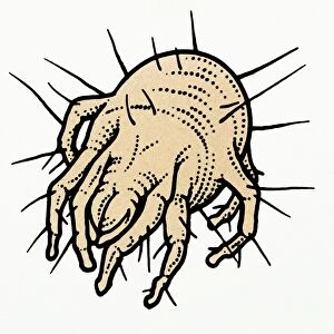
Dermatophagoides pteronyssius, dust mite

![]()
Wall Art and Photo Gifts from Mary Evans Picture Library
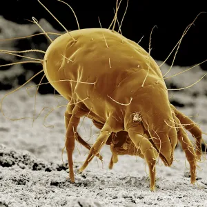
Dermatophagoides pteronyssius, dust mite
Scanning electron microscope image showing a dust mite (x 250 on standard 9cm wide print). This image has been artificially coloured by a computer
Mary Evans Picture Library makes available wonderful images created for people to enjoy over the centuries
Media ID 8580293
© Mary Evans Picture Library 2015 - https://copyrighthub.org/s0/hub1/creation/maryevans/MaryEvansPictureID/10708114
Acari Acarina Arachnid Arachnida Arthropod Arthropoda Dust Electron Electron Micrograph Micrograph Microscope Microscope Image Mite Scanning Scanning Electron Micrograph Scanning Electron Microscope Scanning Electron Microscope Image Dermatophagoides Dermatophagoides Pteronyssinus Dust Mite House Dust Mite Invertebrata
EDITORS COMMENTS
1. Title: A Closer Look into the Microcosm: Dermatophagoides pteronyssius, the Unseen Resident of Our Homes (Scanning Electron Microscope Image) This scanning electron microscope image reveals the intricate structure of Dermatophagoides pteronyssius, commonly known as the house dust mite. With a magnification of 250x on a standard 9cm wide print, this micrograph provides an up-close and colorful glimpse into the world of these microscopic arthropods. Dermatophagoides pteronyssius is an invertebrate belonging to the acariformes order, specifically the pyroglyphidae family. This arachnid, a member of the acari class, is a common inhabitant of household dust. Despite their tiny size, they play a significant role in indoor allergens that can trigger allergic reactions and asthma symptoms in humans. The scanning electron microscope image showcases the dust mite's unique features, including its eight legs, two-segmented antennae, and a pair of chelicerae, which are modified appendages used for grasping and piercing. The micrograph also reveals the dust mite's cuticle, a protective layer that covers its exoskeleton, and its numerous setae, tiny hairs that help it move and sense its environment. The artificially colored image, produced by a computer, enhances the visual appeal and highlights the intricate details of the dust mite's structure. This striking micrograph serves as a reminder of the vast and diverse microscopic life that exists in our homes, often unseen to the naked eye. In summary, this scanning electron microscope image of Dermatophagoides pteronyssius offers a fascinating glimpse into the hidden world of house dust mites, revealing their complex structure and shedding light on their role in indoor allergens.
MADE IN THE USA
Safe Shipping with 30 Day Money Back Guarantee
FREE PERSONALISATION*
We are proud to offer a range of customisation features including Personalised Captions, Color Filters and Picture Zoom Tools
SECURE PAYMENTS
We happily accept a wide range of payment options so you can pay for the things you need in the way that is most convenient for you
* Options may vary by product and licensing agreement. Zoomed Pictures can be adjusted in the Cart.