Fraud Blocker Skip to main content

Scanning Electron Microscope Collection

"Unveiling the Hidden World

Background imageScanning Electron Microscope Collection: X and Y chromosomes

X and Y chromosomes, coloured scanning electron micrograph (SEM). Humans have 46 chromosomes in total: 23 inherited from the mother and 23 from the father

Background imageScanning Electron Microscope Collection: Lavender pollen grain, SEM

Lavender pollen grain, SEM
Lavender pollen grain. Coloured scanning electron micrograph (SEM) of a pollen grain (yellow) amongst the petal (papilla) cells (purple) of a French lavender (Lavandula dentata) flower

Background imageScanning Electron Microscope Collection: Scanning Electron Micrograph (SEM): Tardigrade or ‘Water Bear Magnification x 1250 (A4 size: 29)

Scanning Electron Micrograph (SEM): Tardigrade or ‘Water Bear Magnification x 1250 (A4 size: 29. 7 cm width)
LRDS-129 Tardigrade or Water Bear Scanning Electron Micrograph (SEM) Ramazzotius oberhauseri Magnification x 1250 (A4 size: 29.7 cm width) Coloured by hand to enhance natural features

Background imageScanning Electron Microscope Collection: Cat tongue surface, SEM

Cat tongue surface, SEM
Cats tongue. Coloured scanning electron micrograph (SEM) of the surface of a cats (family Felidae) tongue. The tongue is covered in many backward facing papillae (projections)

Background imageScanning Electron Microscope Collection: Nerve cell, SEM

Nerve cell, SEM
Nerve cell. Coloured scanning electron micrograph (SEM) of a nerve cell (neuron). Neurons are responsible for passing information around the central nervous system (CNS)

Background imageScanning Electron Microscope Collection: Tardigrade or Water Bear

Tardigrade or Water Bear
Water bear or tardigrade. Scanning electron micrograph (SEM) of a water bear (Echiniscus sp.) walking over spagnum moss. Water bears live in damp habitats such as moss or lichen

Background imageScanning Electron Microscope Collection: Scanning Electron Micrograph (SEM): Praying Mantis - Magnification x 30 (if print A4 size: 29)

Scanning Electron Micrograph (SEM): Praying Mantis - Magnification x 30 (if print A4 size: 29. 7 cm wide)
LRDS-392 Scanning Electron Micrograph (SEM): Praying Mantis Magnification x 30 (if print A4 size: 29.7 cm wide) Mantis religiosa David Spears (Last Refuge)

Background imageScanning Electron Microscope Collection: T lymphocytes and cancer cell, SEM C001 / 1679

T lymphocytes and cancer cell, SEM C001 / 1679
T lymphocytes and cancer cell. Coloured scanning electron micrograph (SEM) of T lymphocyte cells (green) attached to a cancer cell

Background imageScanning Electron Microscope Collection: Fruit fly, SEM Z340 / 0768

Fruit fly, SEM Z340 / 0768
Fruit fly. Coloured scanning electron micrograph (SEM) of a fruit fly (Drosophila funebris) on an apple. Its compound eyes (red) are seen and its wings are outstretched

Background imageScanning Electron Microscope Collection: Crysotile asbestos

Crysotile asbestos
Scanning electron micrograph of 5-Fold symmetry in crysotile asbestos. Magnification on the 5 x4 transparency = X 600, 000

Background imageScanning Electron Microscope Collection: Uterus lining during menstruation, SEM

Uterus lining during menstruation, SEM
Uterus during menstruation. Coloured scanning electron micrograph (SEM) of the lining of the uterus being shed during menstruation

Background imageScanning Electron Microscope Collection: Vascular bundle, SEM

Vascular bundle, SEM
Vascular bundle. Coloured scanning electron micrograph (SEM) of a section through a vascular bundle from a rootlet of a dicotyledon plant

Background imageScanning Electron Microscope Collection: Geranium anther, SEM

Geranium anther, SEM
Geranium anther. Coloured scanning electron micrograph (SEM) of pollen (pink) on the anther of a geranium flower (Geranium sp.)

Background imageScanning Electron Microscope Collection: Dahlia flower pollen, SEM

Dahlia flower pollen, SEM
Dahlia flower pollen. Coloured scanning electron micrograph (SEM) of a pollen grain (orange) from a Dahlia cultorum flower. A pollen grain contains a male sex cell from a flowering plant

Background imageScanning Electron Microscope Collection: Botanik Digitalis purpurea L. Fingerhut 160: 1

Botanik Digitalis purpurea L. Fingerhut 160: 1
Foxglove leaf. Coloured scanning electron micrograph (SEM) of a section through a leaf from a foxglove (Digitalis purpurea) plant, showing its hairs (trichomes)

Background imageScanning Electron Microscope Collection: Neutrophil engulfing MRSA, SEM C018 / 8596

Neutrophil engulfing MRSA, SEM C018 / 8596
Neutrophil engulfing MRSA. Coloured scanning electron micrograph (SEM) of a neutrophil white blood cell (green) engulfing methicillin-resistant Staphylococcus aureus bacteria (MRSA, pink)

Background imageScanning Electron Microscope Collection: Passion flower pollen, SEM

Passion flower pollen, SEM
Passion flower pollen. Coloured scanning electron micrograph (SEM) of a passion flower (Passiflora caerulea) pollen grain. Pollen grains are the male gametes (sex cells) of a plant

Background imageScanning Electron Microscope Collection: Liver

Liver
Scanning electron microscope (SEM) image of a section through a liver (x 7000), an organ that has over 500 functions in the human body (x 800)

Background imageScanning Electron Microscope Collection: Cimex lectularius, bed bug

Cimex lectularius, bed bug
Scanning electron microscope image of a bed bug (x 17). The sucking mouthparts enable the feeding bedbugs to pierce the hosts tissues and siphon out a blood meal

Background imageScanning Electron Microscope Collection: Head louse, SEM

Head louse, SEM
Head louse. Coloured scanning electron micrograph (SEM) of a head louse (Pediculus humanus capitis) clinging to a human hair. Head lice measure 2-3 millimetres in length

Background imageScanning Electron Microscope Collection: Acrosphaera radiolarian, SEM

Acrosphaera radiolarian, SEM
Acrosphaera radiolarian. Coloured scanning electron micrograph (SEM) of the shell of a Acrosphaera sp. radiolarian. Radiolaria are single-celled protozoans that are found in marine plankton

Background imageScanning Electron Microscope Collection: Dog tapeworm head, SEM

Dog tapeworm head, SEM
Dog tapeworm head. Coloured scanning electron micrograph (SEM) of the head (scolex) of a dog tapeworm (Taenia pisiformis)

Background imageScanning Electron Microscope Collection: Ant, SEM

Ant, SEM
Ant. Coloured scanning electron micrograph (SEM) of an ant (family Formicidae). One of its compound eyes (red) is seen, as well as two long antennae that are mounted on the head between the eyes

Background imageScanning Electron Microscope Collection: Foot of a chameleon

Foot of a chameleon. Coloured scanning electron micrograph (SEM) of the foot of a chameleon. Chameleons are the only lizards with zygodactyle feet, or with toes in a pincer arrangement

Background imageScanning Electron Microscope Collection: Woven fabric, SEM

Woven fabric, SEM
Woven fabric fibres. Coloured scanning electron micrograph (SEM) of synthetic fibres woven to form clothes. Nylon and polyester are common synthetic clothing materials

Background imageScanning Electron Microscope Collection: Brain surface, SEM

Brain surface, SEM
Brain surface. Coloured scanning electron micrograph (SEM) of the surface of a brain ventricle. Ventricles are cavities within the brain that are continuous with the central canal of the spinal cord

Background imageScanning Electron Microscope Collection: Pollen grains, SEM

Pollen grains, SEM
Pollen grains. Coloured scanning electron micrographs (SEM) of pollen grains from a variety of plants

Background imageScanning Electron Microscope Collection: Leaf pores, SEM

Leaf pores, SEM
Leaf pores. Coloured scanning electron micrograph (SEM) of stomata (holes) on the surface of a leaf. These structures perform a similar function to the pores in human skin

Background imageScanning Electron Microscope Collection: Salt grains and ground peppercorn, SEM

Salt grains and ground peppercorn, SEM
Salt grains and ground peppercorn, coloured scanning electron micrograph (SEM). Each salt (sodium chloride) crystal (white) is composed of a cubic lattice of sodium and chloride ions

Background imageScanning Electron Microscope Collection: Diatom, SEM

Diatom, SEM
Diatom. Coloured scanning electron micrograph (SEM) of the Arachnoidiscus sp. diatom. The diatoms are a group of photosynthetic, single- celled algae containing about 10, 000 species

Background imageScanning Electron Microscope Collection: Diatom, SEM

Diatom, SEM
Diatom. Coloured scanning electron micrograph (SEM) of a Stephanopyxis sp. diatom. Diatoms are single-celled photosynthetic algae, of which there are about 100, 000 species

Background imageScanning Electron Microscope Collection: E. coli bacteria, SEM

E. coli bacteria, SEM
E. coli bacteria. Coloured scanning electron micrograph (SEM) of Escherichia coli bacteria. These bacteria are a normal part of the intestinal flora in humans and other animals

Background imageScanning Electron Microscope Collection: Diatom, SEM

Diatom, SEM
Diatom. Coloured scanning electron micrograph (SEM) of a Triceratium morlandii diatom. Diatoms are single-celled photosynthetic algae, of which there are about 100, 000 species

Background imageScanning Electron Microscope Collection: Scanning Electron Micrograph (SEM): Fruit Fly, Magnification x 300 (A4 size: 29. 7 cm width)

Scanning Electron Micrograph (SEM): Fruit Fly, Magnification x 300 (A4 size: 29. 7 cm width)
LRDS-57 Fruit Fly Scanning Electron Micrograph (SEM) Drosophila sp Magnification x 300 (A4 size: 29.7 cm width) Coloured by hand to enhance natural features

Background imageScanning Electron Microscope Collection: Culex mosquito, SEM

Culex mosquito, SEM
Culex mosquito. Coloured scanning electron micrograph (SEM) of a Culex sp. mosquito. The mosquitos head is dominated by its large compound eyes (brown spheres)

Background imageScanning Electron Microscope Collection: Red-barbed ant, SEM

Red-barbed ant, SEM
Red-barbed ant (Formica rufibarbis), coloured scanning electron micrograph (SEM). This ant is common in mainland Europe, but rare in the UK

Background imageScanning Electron Microscope Collection: Embryonic stem cell and needle, SEM

Embryonic stem cell and needle, SEM
Embryonic stem cell and needle. Coloured scanning electron micrograph (SEM) of an embryonic stem cell (ESC) sitting in the eye of a needle

Background imageScanning Electron Microscope Collection: Calcareous phytoplankton, SEM

Calcareous phytoplankton, SEM
Calcareous phytoplankton. Coloured scanning electron micrograph (SEM) of the external mineralised structures (coccospheres) of small marine algal organisms called coccolithophores

Background imageScanning Electron Microscope Collection: Salmonella bacteria, SEM

Salmonella bacteria, SEM
Salmonella bacteria, coloured scanning electron micrograph (SEM). These Gram-negative rod-shaped bacteria can cause food poisoning when eaten in contaminated food

Background imageScanning Electron Microscope Collection: Candida fungus, SEM

Candida fungus, SEM
Candida fungus. Coloured scanning electron micrograph (SEM) of budding threads (hyphae) of a Candida fungus. The budding areas are where asexual reproduction is producing new fungus cells

Background imageScanning Electron Microscope Collection: Taraxacum officinale, dandelion (fruiting head)

Taraxacum officinale, dandelion (fruiting head)
Scanning electron microscope image showing a vertical section through an unripe fruiting head of a dandelion in the yellow flower stage. Colour added artificially by computer

Background imageScanning Electron Microscope Collection: Sheep tick, SEM

Sheep tick, SEM
Sheep tick. Coloured scanning electron micrograph (SEM) of a sheep tick (Ixodes ricinus). The ticks mouthparts are between its two front legs

Background imageScanning Electron Microscope Collection: Small intestine, SEM

Small intestine, SEM
Small intestine. Coloured scanning electron micrograph (SEM) of a biopsy from the small intestines of a child. The small intestine runs from the stomach to the large intestine

Background imageScanning Electron Microscope Collection: Gallbladder surface, SEM

Gallbladder surface, SEM
Gallbladder. Coloured scanning electron micrograph (SEM) of the surface of the gallbladder showing the numerous microvilli that cover the columnar epithelium

Background imageScanning Electron Microscope Collection: Moth proboscis, SEM

Moth proboscis, SEM
Moth proboscis. Coloured scanning electron micrograph (SEM) of the coiled proboscis of a moth (order Lepidoptera). The proboscis is an elongated part of the mouth

Background imageScanning Electron Microscope Collection: Honey bee, SEM

Honey bee, SEM
Honey bee (Apis mellifera), coloured scanning electron micrograph (SEM). The head and one of the two compound eyes is at lower left

Background imageScanning Electron Microscope Collection: Honeybee leg, SEM

Honeybee leg, SEM
Honey bee leg. Coloured scanning electron micrograph (SEM) of the leg of a honeybee (Apis mellifera) covered in pollen grains (round). The dense coating of hairs has trapped the pollen

Background imageScanning Electron Microscope Collection: Head of a honey bee, SEM

Head of a honey bee, SEM
Head of a honey bee (Apis mellifera), coloured scanning electron micrograph (SEM). The compound eyes (one seen) are either side of the antennae


All Professionally Made to Order for Quick Shipping

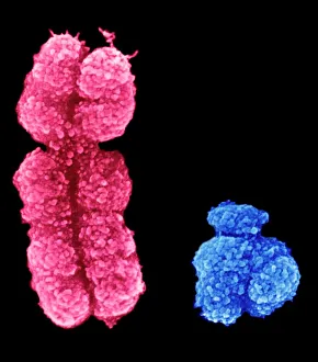
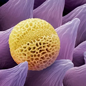
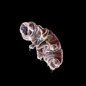
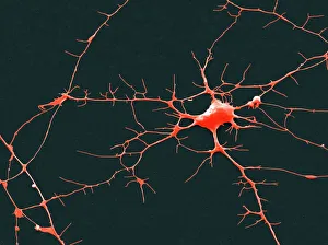
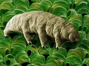
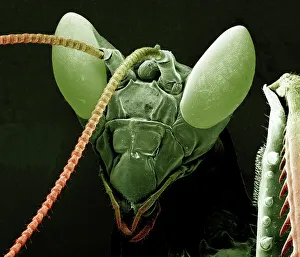
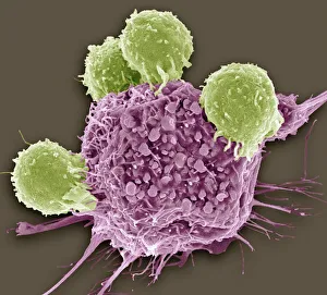
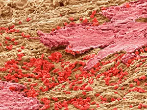
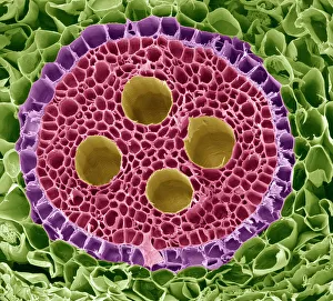
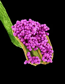
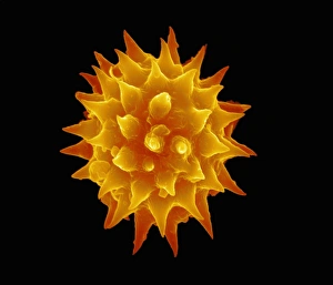
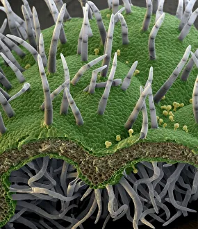
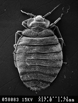
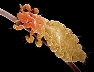
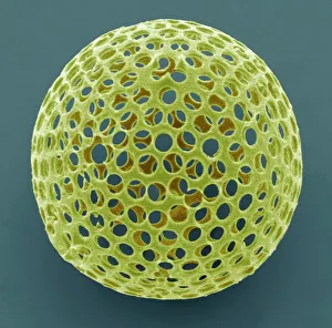
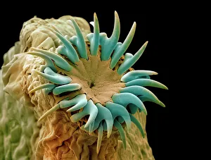
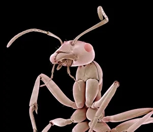
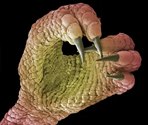
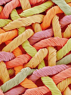
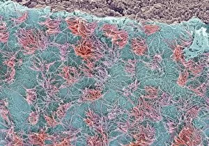
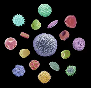
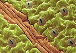
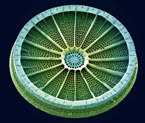
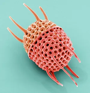
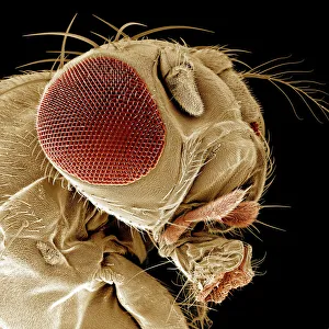
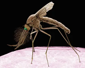
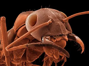
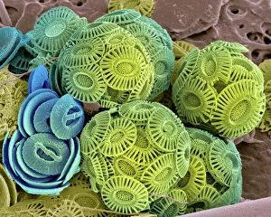
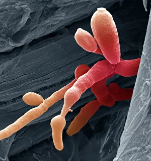
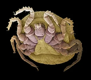
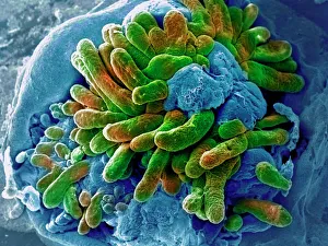
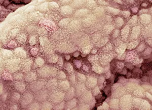
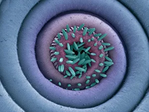
"Unveiling the Hidden World: Exploring with a Scanning Electron Microscope" The scanning electron microscope (SEM) is an incredible tool that allows us to delve into the microscopic realm, revealing intricate details and unlocking secrets of the unseen. With its high magnification capabilities, it offers a glimpse into fascinating aspects of life and nature. In one captivating image, we witness the beauty of gender determination as X and Y chromosomes are captured under the SEM's lens. The intricate patterns and structures within these chromosomes showcase the complexity of our genetic makeup. Moving on to another astonishing discovery, a Scanning Electron Micrograph presents us with an up-close view of a Praying Mantis at 30 times magnification. Every tiny detail becomes visible, from its delicate limbs to its mesmerizing eyes – reminding us of nature's remarkable diversity. Shifting focus to plant life, lavender pollen grains come alive in another SEM image. These minuscule particles take center stage as their unique shapes and textures become apparent under intense scrutiny. Zooming in even further reveals a Tardigrade or 'Water Bear' at an astounding 1250 times magnification. This resilient creature captures our imagination as we explore its alien-like features through this powerful imaging technique. Delving deeper into biological wonders, T lymphocytes battling cancer cells are showcased in yet another breathtaking SEM capture. This visual representation highlights the ongoing fight within our bodies against diseases like cancer – emphasizing both hope and resilience. Calcareous phytoplankton takes center stage next as we uncover their intricate forms through SEM imagery. These microscopic organisms play crucial roles in marine ecosystems while leaving us awe-inspired by their stunningly detailed structures. Transitioning to neural marvels, nerve cells reveal their intricacy under the watchful eye of an SEM. Their branching extensions create complex networks that enable communication throughout our bodies – showcasing nature's ingenuity at work. Venturing into insect realms once again, a fruit fly is immortalized under the SEM's lens.