Home > Animals > Worms > Flukes
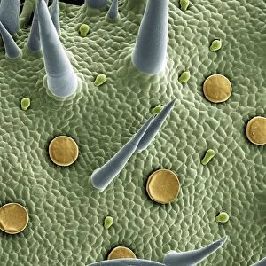
Schistosoma nasale, bloodfluke

![]()
Wall Art and Photo Gifts from Mary Evans Picture Library
Schistosoma nasale, bloodfluke
Scanning electron microscope image of a parasitic bloodfluke or flatworm. Coloured artifically by computer
Mary Evans Picture Library makes available wonderful images created for people to enjoy over the centuries
Media ID 8587905
© Mary Evans Picture Library 2015 - https://copyrighthub.org/s0/hub1/creation/maryevans/MaryEvansPictureID/10712997
Alex Alex Ball Blood Chris Chris Jones Disease Electron Electron Micrograph Flatworm Fluke Jones Mammalia Micrograph Microscope Microscope Image Parasite Bilharzia Invertebrata
EDITORS COMMENTS
1. Title: A Closer Look into the Microcosm: Schistosoma nasale, the Blood-Sucking Parasitic Flatworm Schistosoma nasale, a blood-dwelling flatworm and a notorious parasite, is the focus of this scanning electron microscope image. This parasitic invertebrate, belonging to the Digenean class of flatworms, is responsible for causing diseases in mammals, including humans, leading to conditions such as schistosomiasis or bilharzia. In this intricately detailed micrograph, the parasite's complex morphology is brought to life through artificial coloring by computer. The image reveals the various structures of the flatworm, including its tegument, which is covered in tiny, overlapping scales, and its suckers, designed for attachment to the host's blood vessels. The presence of Schistosoma nasale in the bloodstream of its mammalian host can lead to a range of symptoms, including fever, anemia, and intestinal or urinary tract damage. The parasite's life cycle involves multiple hosts, including snails, which act as intermediate hosts, and the final mammalian host, where the parasite matures and lays eggs. This micrograph offers a unique perspective into the intricate world of parasitic flatworms and the complex relationships they form with their hosts. It serves as a reminder of the vast and diverse array of organisms that inhabit our world, many of which remain hidden from our everyday view. Alex Ball, a renowned microscopist, and Chris Jones, a skilled technician, collaborated on capturing this stunning image, using advanced scanning electron microscopy techniques to bring the hidden world of Schistosoma nasale to light. This image is an essential addition to any collection of micrographs, showcasing the beauty and complexity of the natural world.
MADE IN THE USA
Safe Shipping with 30 Day Money Back Guarantee
FREE PERSONALISATION*
We are proud to offer a range of customisation features including Personalised Captions, Color Filters and Picture Zoom Tools
SECURE PAYMENTS
We happily accept a wide range of payment options so you can pay for the things you need in the way that is most convenient for you
* Options may vary by product and licensing agreement. Zoomed Pictures can be adjusted in the Cart.