
Microscope Image Collection (page 3)
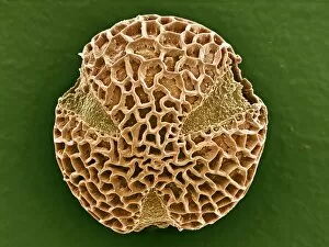
"Unlocking the Hidden World: A Glimpse through the Microscope" Delving into the intricate beauty of nature, this microscope image reveals Discosphaera tubifera


All Professionally Made to Order for Quick Shipping
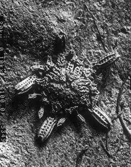
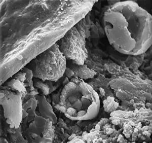
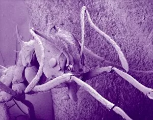
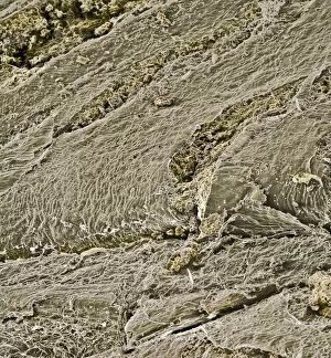
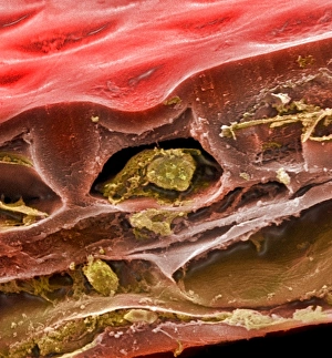
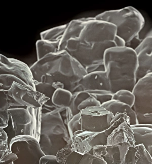
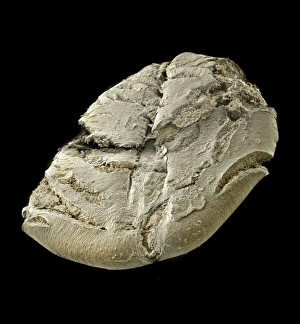
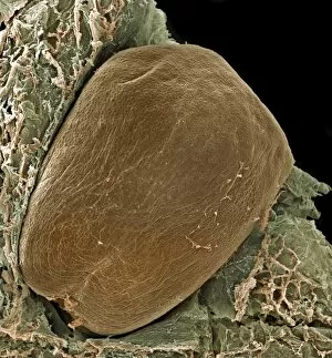
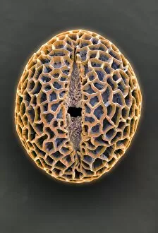
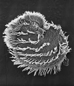
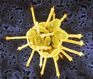
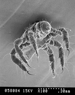
"Unlocking the Hidden World: A Glimpse through the Microscope" Delving into the intricate beauty of nature, this microscope image reveals Discosphaera tubifera, a mesmerizing coccolithophore that adorns our oceans with its delicate structure. Exploring beyond what meets the eye, this microscopic view uncovers Crysotile asbestos fibers, highlighting their hazardous presence and emphasizing the need for caution in industrial settings. Peering into the depths of life's vital organ, we witness liver cells in astonishing detail, marveling at their complex network and crucial role in maintaining our well-being. Zooming in on an unexpected intruder within our homes, this close-up captures Cimex lectularius - commonly known as bed bugs - reminding us to remain vigilant against these resilient pests. Nature's resilience takes center stage as we examine Taraxacum officinale's fruiting head under a microscope; dandelion seeds ready to disperse and conquer new territories with their remarkable adaptability. Unveiling nature's hidden weaponry, this fascinating image showcases snail teeth - tiny yet powerful structures designed for devouring tough vegetation and leaving behind intricate patterns on surfaces they graze upon. Diving into the realm of disease-causing organisms, we encounter Plasmodium sp. , a malarial parasite responsible for countless human suffering worldwide; a stark reminder of ongoing medical challenges faced by humanity. Shining light on minerals' captivating allure, kaolinite crystals reveal themselves through this microscope lens – showcasing their unique arrangement and contributing to various industries from ceramics to cosmetics. Exposing an unwelcome guest beneath our skin’s surface is Sarcoptes scabiei – better known as scabies mites – causing intense itching while teaching us about personal hygiene practices necessary for prevention.