Fraud Blocker Skip to main content

Microscope Image Collection (page 6)

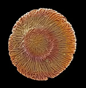
"Unlocking the Hidden World: A Glimpse through the Microscope" Delving into the intricate beauty of nature, this microscope image reveals Discosphaera tubifera

Background imageMicroscope Image Collection: Sugar grains

Sugar grains
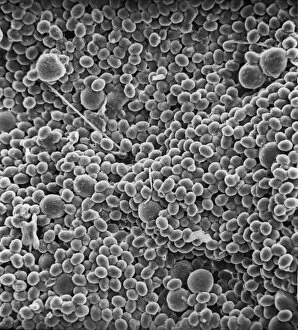
A scanning electron microscope (SEM) image of sugar grains, artificially coloured by computer

Background imageMicroscope Image Collection: Vitis sp. white grape

Vitis sp. white grape
A scanning electron microscope (SEM) image of a white grape (Vitis sp.), artificially coloured by computer

Background imageMicroscope Image Collection: Browallia speciosa, amethyst

Browallia speciosa, amethyst
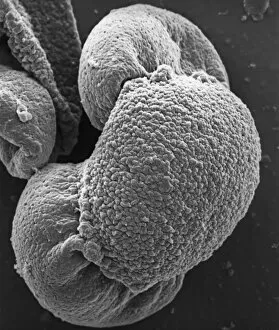
A pollen grain of the Browallia speciosa (polar view) from the family Solanaceae, the tomato family

Background imageMicroscope Image Collection: Vaccinium sp. blueberry

Vaccinium sp. blueberry
A scanning electron microscope (SEM) image of a blueberry (Vaccinium sp.), artificially coloured by computer

Background imageMicroscope Image Collection: Solanum sp. tomato

Solanum sp. tomato
A scanning electron microscope (SEM) image of a tomato (Solanum sp.), artificially coloured by computer

Background imageMicroscope Image Collection: Isurus oxyrinchus, mako shark

Isurus oxyrinchus, mako shark
Scanning Electron Microscope image of mako shark skin

Background imageMicroscope Image Collection: Lasius niger, black garden ant

Lasius niger, black garden ant
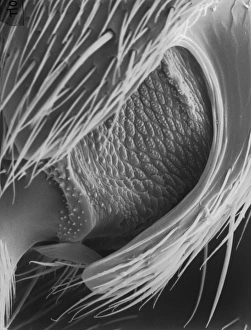
Scanning electron microscope (SEM) of a black ant leg. Widespread and common in a range of habitats but perhaps most familiar in gardens where nests are formed under paving stones and brickwork

Background imageMicroscope Image Collection: Porcellio sccaber, woodlouse

Porcellio sccaber, woodlouse
Scanning electron microscope (SEM) image showing all the units that make up the compound eye of a woodlouse

Background imageMicroscope Image Collection: Calliphora vicina, blowfly or bluebottle

Calliphora vicina, blowfly or bluebottle
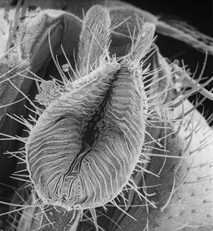
Scanning electron microscope (SEM) image of a blowfly proboscis (x 85). This specialised mouth-part is used to squirt digestive enzymes onto the food

Background imageMicroscope Image Collection: Papilio machaon, old world swallowtail

Papilio machaon, old world swallowtail
SEM image of a Papilio machaon wing

Background imageMicroscope Image Collection: Microscope image of the Johnstown diogenite

Microscope image of the Johnstown diogenite. Diogenites are coarse grained and composed primarily of one mineral, pyroxene. Field of view is 2.5mm across

Background imageMicroscope Image Collection: Microscope image of chondrite

Microscope image of chondrite showing chondrules, typical of primitive meteorites. Horizontal field of view, 3.3mm

Background imageMicroscope Image Collection: Microscope image of the Zagami shergottite

Microscope image of the Zagami shergottite. The fractures in the pyroxene mineral grains and the paler patches of glass show that the rock has been shocked. Field of view is 5mm

Background imageMicroscope Image Collection: Microscope image of the Brachina meteorite

Microscope image of the Brachina meteorite, the type specimen of the Brachinite meteorites. Brachinites are composed mostly of olivine with minor amounts of pyroxene and plagioclase

Background imageMicroscope Image Collection: Microscope image of the Lodran meteorite

Microscope image of the Lodran meteorite. This meteorite is the type specimen of the Lodranite meteorites. The lodranites are related to the acaplucoites but are more course-grained

Background imageMicroscope Image Collection: Optical microscope image of the Barwell (Type 6) chondrite

Optical microscope image of the Barwell (Type 6) chondrite. This meteorite has experienced a significant amount of heating

Background imageMicroscope Image Collection: Optical microscope image of the Parnallee (Type 3) chondrite

Optical microscope image of the Parnallee (Type 3) chondrite
An optical microscope image of the Parnallee (Type 3) chondrite that has experienced little heating. The chondrules are clear and well-defined. The field of view is 5mm

Background imageMicroscope Image Collection: Danionella dracula, minnow

Danionella dracula, minnow
SEM image of the Danionella dracula. This tiny 17mm fish has evolved many unique and unusual characteristics, the most spectacular of which are jaw modifications that resemble true teeth

Background imageMicroscope Image Collection: Feather detail

Feather detail

Background imageMicroscope Image Collection: Pollen on bee

Pollen on bee
Scanning electron microscope (SEM) image of pollen on a bee. If the plant depends on animals for pollination, the pollen will be relatively large and sticky

Background imageMicroscope Image Collection: Dermestes lardarius, larder beetle

Dermestes lardarius, larder beetle
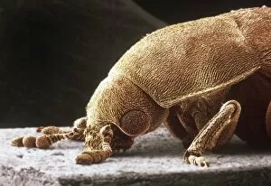
Scanning electron microscope image of a larder beetle (x22). These beetles are important for the damage they do, mainly through feeding on animal matter. Coloured artificially by computer

Background imageMicroscope Image Collection: Calcidiscus leptoporus and Syracolithus quadriperforatus, co

Calcidiscus leptoporus and Syracolithus quadriperforatus, co
In this scanning electron micrograph, the transition of a life-cycle stage in Calcidiscus is shown from the outer cover to the inner layer. Specimen taken from W. Mediterranean

Background imageMicroscope Image Collection: Apis mellifera, honey bee

Apis mellifera, honey bee
Scanning electron microscope image of a honey bee coloured artificially by computer. The female worker caste of this species have special baskets on their legs to to take pollen back to the nest

Background imageMicroscope Image Collection: Actinopora disticha, bryozoan

Actinopora disticha, bryozoan
Scanning electron micrograph of a fossil cyclostome bryozoan from the Cretaceous Chalk, Santonian, Kent

Background imageMicroscope Image Collection: Wilbertopora woodwardi (Brydone), bryozoan

Wilbertopora woodwardi (Brydone), bryozoan
Scanning electron micrograph of a fossil cheilostome bryozoan. Specimen originates from the Upper Cretaceous Chalk, West Mean Station, Hampshire, U.K

Background imageMicroscope Image Collection: Ptinus tectus, spider beetle

Ptinus tectus, spider beetle
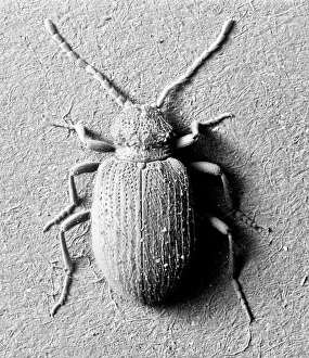
Scanning electron microscope image of a spider beetle (x 9). The long antennae, hairy body and waist-like constriction give this beetle the appearance of a spider

Background imageMicroscope Image Collection: Aspidelectra melolontha, bryozoan

Aspidelectra melolontha, bryozoan
Scanning electron micrograph. Zooids of a bleached colony of a modern cheilostome bryozoan. A recent specimen from Sheppey, Kent

Background imageMicroscope Image Collection: Pinus sylvestris, scots pine

Pinus sylvestris, scots pine
Scanning electron microscope (SEM) image showing a pollen grain from a scots pine. Note the air bladders that help it to float through the air (x 1500 on a standard 9 cm wide print)

Background imageMicroscope Image Collection: Actinoptychus, diatom

Actinoptychus, diatom
Scanning electron microscope image of the exterior valve of the diatom Actinoptychus (x 500 on a standard 9 cm wide print)

Background imageMicroscope Image Collection: Chenopodium album, goosefoot

Chenopodium album, goosefoot
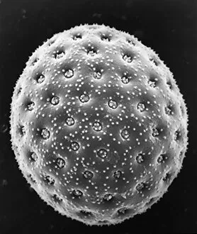
Scanning electron microscope image of a pollen grain from a member of the goosefoot family (x 3000 on a standard 9 cm wide print)

Background imageMicroscope Image Collection: Pthirus gorillae, gorilla louse

Pthirus gorillae, gorilla louse
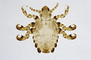
Photomicrograph of a gorilla louse specimen, length 2.5 mm from Rwanda / Zaire. The gorilla louse is from the same genus as the human louse

Background imageMicroscope Image Collection: Gymnosperm, palm & angiosperm

Gymnosperm, palm & angiosperm
Composite photomicrograph comparing structures visible through a hand-held lens in transverse section of fossil tree trunks: gymnosperm (left), palm (centre) and angiosperm (right)

Background imageMicroscope Image Collection: Granite from Ailsa Craig

Granite from Ailsa Craig
A photomicrograph of granite taken between crossed polarisers. Granite is an igneous rock


All Professionally Made to Order for Quick Shipping

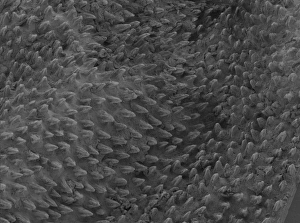
"Unlocking the Hidden World: A Glimpse through the Microscope" Delving into the intricate beauty of nature, this microscope image reveals Discosphaera tubifera, a mesmerizing coccolithophore that adorns our oceans with its delicate structure. Exploring beyond what meets the eye, this microscopic view uncovers Crysotile asbestos fibers, highlighting their hazardous presence and emphasizing the need for caution in industrial settings. Peering into the depths of life's vital organ, we witness liver cells in astonishing detail, marveling at their complex network and crucial role in maintaining our well-being. Zooming in on an unexpected intruder within our homes, this close-up captures Cimex lectularius - commonly known as bed bugs - reminding us to remain vigilant against these resilient pests. Nature's resilience takes center stage as we examine Taraxacum officinale's fruiting head under a microscope; dandelion seeds ready to disperse and conquer new territories with their remarkable adaptability. Unveiling nature's hidden weaponry, this fascinating image showcases snail teeth - tiny yet powerful structures designed for devouring tough vegetation and leaving behind intricate patterns on surfaces they graze upon. Diving into the realm of disease-causing organisms, we encounter Plasmodium sp. , a malarial parasite responsible for countless human suffering worldwide; a stark reminder of ongoing medical challenges faced by humanity. Shining light on minerals' captivating allure, kaolinite crystals reveal themselves through this microscope lens – showcasing their unique arrangement and contributing to various industries from ceramics to cosmetics. Exposing an unwelcome guest beneath our skin’s surface is Sarcoptes scabiei – better known as scabies mites – causing intense itching while teaching us about personal hygiene practices necessary for prevention.