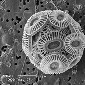
Amphitetras, diatom

![]()
Wall Art and Photo Gifts from Mary Evans Picture Library
Amphitetras, diatom
Scanning electron microscope (SEM) image showing the diatom Amphitetras with its ornate silica shell (x5000 on a standard 9 cm wide print). Coloured artificially by computer
Mary Evans Picture Library makes available wonderful images created for people to enjoy over the centuries
Media ID 8609450
© Mary Evans Picture Library 2015 - https://copyrighthub.org/s0/hub1/creation/maryevans/MaryEvansPictureID/10715279
Alga Algae Algal Bacillariophyceae Chromalveolata Chromista Diatom Electron Electron Micrograph Eukaryote Eukaryotic Magnification Micrograph Microscope Microscope Image Ochrophyta Protist Protista Scanning Scanning Electron Micrograph Scanning Electron Microscope Scanning Electron Microscope Image Sem Image
EDITORS COMMENTS
1. Title: "Amphitetras: A Magnificent Diatom Revealed through Scanning Electron Microscopy" Amphitetras, a captivating diatom from the family Coscinodiscophycidae within the order Coscinodiscophyceae, is showcased in this stunning Scanning Electron Microscope (SEM) image. Diatoms, a major group of algae belonging to the phylum Bacillariophyceae, are renowned for their intricately patterned silica shells, which provide both structural support and protection. In this high-magnification SEM image, Amphitetras is depicted at x5000 magnification on a standard 9 cm wide print. The diatom's ornate shell is coloured artificially by computer to enhance its visual appeal. The silica shell is composed of two distinct halves, which are intricately interconnected, forming a beautiful, symmetrical structure. The central part of the shell, known as the epitheca, is adorned with a complex, labyrinthine pattern, while the valve flange, or the rim of the shell, exhibits a more uniform, smooth texture. Amphitetras is a member of the Chromalveolata, a large and diverse group of eukaryotes that includes algae, protists, and other organisms. This diatom's intricate design is a testament to the remarkable complexity and diversity of life at the microscopic level. The SEM image offers a unique perspective, revealing the intricacies of Amphitetras' shell that would otherwise go unnoticed to the naked eye. This image serves as a reminder of the beauty and complexity of the natural world, and the role that advanced microscopy techniques, such as SEM, play in revealing the hidden wonders that lie beneath our everyday perception.
MADE IN THE USA
Safe Shipping with 30 Day Money Back Guarantee
FREE PERSONALISATION*
We are proud to offer a range of customisation features including Personalised Captions, Color Filters and Picture Zoom Tools
SECURE PAYMENTS
We happily accept a wide range of payment options so you can pay for the things you need in the way that is most convenient for you
* Options may vary by product and licensing agreement. Zoomed Pictures can be adjusted in the Cart.