Fraud Blocker Skip to main content

Magnified Image Collection (page 4)

"Exploring the unseen wonders of the microscopic world: from particle tracks to intricate tissues and beyond

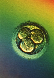
Background imageMagnified Image Collection: LM of four-cell embryo

LM of four-cell embryo
Four-cell embryo. Light micrograph of the blasto- meres of a four-cell human embryo, two days after fertilisation. The blastomeres are the rounded cells

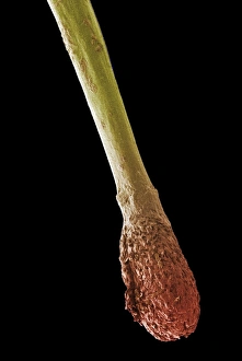
Background imageMagnified Image Collection: Hair root, SEM

Hair root, SEM
Hair root. Coloured scanning electron micrograph (SEM) of the root (red) and part of the shaft (yellow) of a human hair. The base of the hair is known as the hair bulb and is the site of hair growth

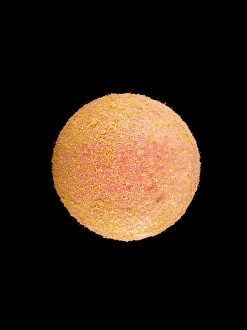
Background imageMagnified Image Collection: Coloured SEM of a fertilized human egg (zygote)

Coloured SEM of a fertilized human egg (zygote)

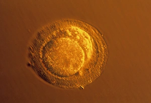
Background imageMagnified Image Collection: Sperm fertilising egg

Sperm fertilising egg

Background imageMagnified Image Collection: LM of blastomeres of a two-cell embryo

LM of blastomeres of a two-cell embryo
Two-cell embryo. Light micrograph of the blasto- meres of a 2-cell human embryo, two days after fertilisation. The blastomeres are the rounded cells formed by the first division of the fertilized

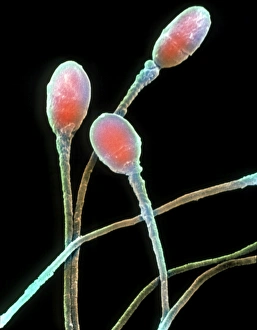
Background imageMagnified Image Collection: Human sperm

Human sperm

Background imageMagnified Image Collection: Mitotic cell division, TEM

Mitotic cell division, TEM
Mitotic cell division. Coloured transmission electron micrograph (TEM) of a cell during the metaphase stage of mitosis (cell division)

Background imageMagnified Image Collection: Periodontal ligament fibres, SEM

Periodontal ligament fibres, SEM
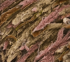
Periodontal ligament fibres. Coloured scanning electron micrograph (SEM) of periodontal ligament fibres (red). These fibres, a specialised type of connective tissue

Background imageMagnified Image Collection: Ovarian tissue, SEM

Ovarian tissue, SEM
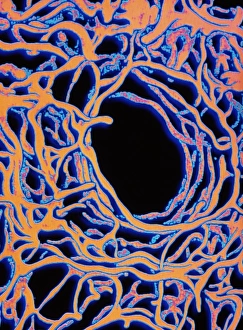
Ovarian tissue. Coloured scanning electron micrograph (SEM) of a section through the medulla of an ovary showing blood vessels (pink)

Background imageMagnified Image Collection: Kidney tubule, TEM

Kidney tubule, TEM
Kidney tubule. Coloured transmission electron micrograph (TEM) of a section through a proximal convoluted tubule in the kidney

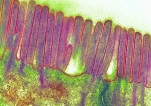
Background imageMagnified Image Collection: Duodenal microvilli

Duodenal microvilli
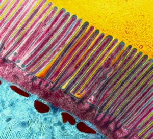
Microvilli in duodenum. Coloured transmission electron micrograph (TEM) of a section through the human duodenum, showing microvilli on the surface epithelium (lining)

Background imageMagnified Image Collection: Coloured TEM of microvilli of intestinal cell

Coloured TEM of microvilli of intestinal cell

Background imageMagnified Image Collection: Liver capillary, SEM

Liver capillary, SEM
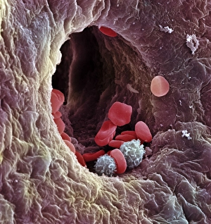
Liver capillary. Coloured scanning electron micro- graph (SEM) of a freeze-fractured section through liver tissue showing a hepatic capillary, or sinusoid

Background imageMagnified Image Collection: Corpus luteum, SEM

Corpus luteum, SEM
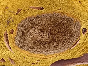
Corpus luteum. Coloured scanning electron micrograph (SEM) of a corpus luteum, or yellow body (brown), in an ovary. The corpus luteum is formed after ovulation

Background imageMagnified Image Collection: Coloured SEM of glomeruli in the human kidney

Coloured SEM of glomeruli in the human kidney

Background imageMagnified Image Collection: Pancreas cell, TEM

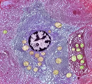
Pancreas cell, TEM
Pancreas cell. Coloured transmission electron micrograph (TEM) of an acinar (exocrine) pancreatic cell. Acinar cells secrete the inactive precursors (zymogens)

Background imageMagnified Image Collection: Kidney glomerulus

Kidney glomerulus
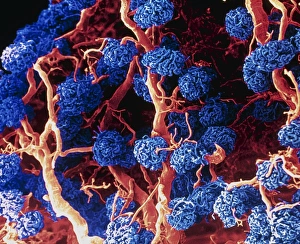
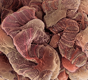
Glomerulus. Coloured scanning electron micrograph (SEM) of a healthy kidney glomerulus. The glomerulus is a tight ball of capillaries where blood passing through the kidneys is filtered

Background imageMagnified Image Collection: Coloured SEM of a sectioned villus from the ileum

Coloured SEM of a sectioned villus from the ileum

Background imageMagnified Image Collection: Kidney podocyte cells, SEM

Kidney podocyte cells, SEM
Kidney podocyte cells. Coloured scanning electron micrograph (SEM) of podocyte cells on the surface of a kidney glomerulus. Podocytes possess branching, foot-like processes (red/brown)

Background imageMagnified Image Collection: Trachea lining, TEM

Trachea lining, TEM
Trachea lining. Coloured transmission electron micrograph (TEM) of a transverse section through the lining of the trachea (windpipe). The trachea links the larynx to the lungs

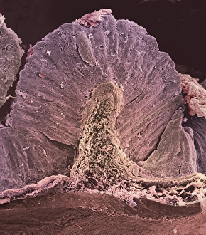
Background imageMagnified Image Collection: False colour SEM of entrance to gastric gland

False colour SEM of entrance to gastric gland
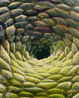
False-colour scanning electron micrograph (SEM) of the mucous membrane lining the stomach, showing the entrance to a gastric gland

Background imageMagnified Image Collection: Stomach wall, SEM

Stomach wall, SEM
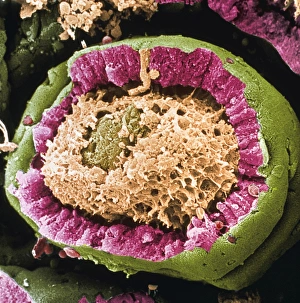
Stomach wall. Coloured scanning electron micrograph (SEM) of a section through a freeze- fractured fold in the stomach wall

Background imageMagnified Image Collection: Ear canal, SEM

Ear canal, SEM
Ear canal. Coloured scanning electron micrograph (SEM) of hairs growing in an ear canal. The ear canal runs between the outer ear and the eardrum

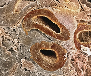
Background imageMagnified Image Collection: Kidney tubules, SEM

Kidney tubules, SEM
Kidney tubules. Coloured scanning electron micrograph (SEM) of proximal convoluted tubules (dark brown) in the kidney. The proximal convoluted tubules function to reabsorb water

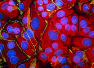
Background imageMagnified Image Collection: Immunofluorescent LM of normal breast cells

Immunofluorescent LM of normal breast cells

Background imageMagnified Image Collection: Inner ear balance sensing, SEM

Inner ear balance sensing, SEM
Inner ear balance sensing. Coloured scanning electron micrograph (SEM) of bundles of cilia (hair cells, yellow) and otoliths (round, red) in the human inner ear

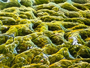
Background imageMagnified Image Collection: False-colour SEM of gastric glands of stomach

False-colour SEM of gastric glands of stomach
Gastric gland: false-colour scanning electron micrograph (SEM) of the mucous membrane lining the stomach, showing the shadowy entrances to a number of gastric glands

Background imageMagnified Image Collection: Rod cells

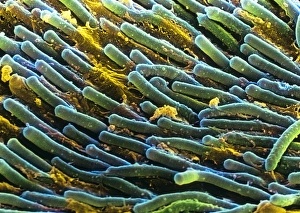
Rod cells. Coloured scanning electron micrograph (SEM) of rod cells (green) in the retina of the eye. Rod cells are light-sensitive cells which respond to dim light

Background imageMagnified Image Collection: Purkinje nerve cells

Purkinje nerve cells

Background imageMagnified Image Collection: Coloured SEM of femoral spongy bone

Coloured SEM of femoral spongy bone

Background imageMagnified Image Collection: Bone, SEM

Bone, SEM
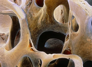
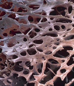
Spongy bone. Coloured scanning electron micrograph (SEM) of freeze-fractured spongy bone tissue. It shows the matrix of collagen and calcium crystals that form the structure of bone tissue

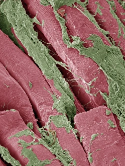
Background imageMagnified Image Collection: Smooth muscle, SEM

Smooth muscle, SEM
Smooth muscle. Coloured scanning electron micrograph of smooth muscle from the trachea (windpipe). Smooth muscle is not under voluntary control, unlike skeletal (striated) muscle

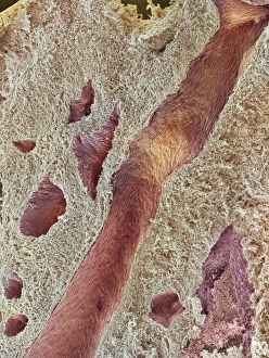
Background imageMagnified Image Collection: Spongy bone, SEM

Spongy bone, SEM
Spongy bone. Coloured scanning electron micrograph (SEM) of human spongy (cancellous) bone from the shaft of a long bone. Found inside bones, below a dense shell of hard compact bone

Background imageMagnified Image Collection: Coloured SEM of section through human spleen

Coloured SEM of section through human spleen
Human spleen. Coloured Scanning Electron Micro- graph (SEM) of a section through the human spleen. Here, the fine network of reticular fibres known as trabeculae is seen


All Professionally Made to Order for Quick Shipping

"Exploring the unseen wonders of the microscopic world: from particle tracks to intricate tissues and beyond. " "Unveiling the hidden beauty within: a closer look at cerebellum tissue through a mesmerizing light micrograph. " "A captivating glimpse into the mysteries of subatomic particles: behold the bubble chamber photo capturing sigma particle decay. " "The groundbreaking moment in science history: witness the first observation of omega-minus particle, forever changing our understanding of matter. " "Awe-inspiring beginnings: marvel at the intricate structure of a human blastocyst, where life takes its first steps. " "Diving deep into neural networks: an illuminating immunofluorescent LM reveals stunning neurons and astrocytes in all their glory. " "Unraveling nature's cycle within us: witness the uterus lining during menstruation, as seen through SEM imaging - a fascinating display of renewal and change. " "Peering into viral threats that loom above us all: explore avian flu virus up close, revealing its complex structure and potential dangers it holds. " "The crystalline beauty behind love and bonding: discover oxytocin hormone crystals under PLM C016 / 7196 microscopy - nature's secret ingredient for connection. " "A tapestry woven by nature itself: delve into intricately detailed fabric structures captured through SEM imaging - artistry on a microscopic scale. " "Glimpsing into windows to our souls with breathtaking precision: explore the iris of an eye like never before, revealing unique patterns that make each person truly one-of-a-kind. " "Revealing fragility beneath strength: examine osteoporotic bone under SEM, shedding light on this silent disease affecting millions worldwide.