Fraud Blocker Skip to main content

Magnified Image Collection (page 12)

"Exploring the unseen wonders of the microscopic world: from particle tracks to intricate tissues and beyond

Background imageMagnified Image Collection: Tongue bacteria, SEM

Tongue bacteria, SEM
Tongue bacteria. Coloured scanning electron micrograph (SEM) of bacteria on the surface of a human tongue. Large numbers of bacteria can form a visible layer on the surface of the tongue

Background imageMagnified Image Collection: Illustration of structures of a typical bacterium

Illustration of structures of a typical bacterium
Cutaway illustration of the internal structure of a typical bacterial cell. This example is of a capsulated bacterium; the capsule (light green) forms the outer of 3 layers

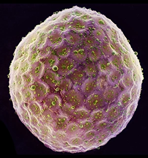
Background imageMagnified Image Collection: Tobacco plant protoplast, SEM

Tobacco plant protoplast, SEM
Protoplast. Coloured scanning electron micrograph (SEM) of a tobacco plant (Nicotiana tabacum) protoplast. A protoplast is a plant cell that has had its tough outer cell wall removed by chemical

Background imageMagnified Image Collection: Salmonella bacterium, computer artwork

Salmonella bacterium, computer artwork. This is a Gram-negative rod-shaped bacteria that has flagella (hair-like structures) that it uses for locomotion

Background imageMagnified Image Collection: Quantum tunneling

Quantum tunneling. Computer model of a quantum wavefunction trapped in a deep well (centre). In classical physics, the particle described by this wavefunction doesn t have enough energy to emerge

Background imageMagnified Image Collection: Female sex hormone

Female sex hormone. Polarised light micrograph of crystals of Oestradiol, also known as beta- Estradiol. This is the most potent of the six naturally-occurring oestrogens

Background imageMagnified Image Collection: Freshwater alga, SEM

Freshwater alga, SEM
Freshwater alga. Coloured scanning electron micrograph (SEM) of a single-celled freshwater alga

Background imageMagnified Image Collection: MRSA bacteria, TEM

MRSA bacteria, TEM
MRSA bacteria. Coloured transmission electron micrograph (TEM) of methicillin-resistant Staphylococcus aureus (MRSA), a gram-positive, round (coccus) bacterium

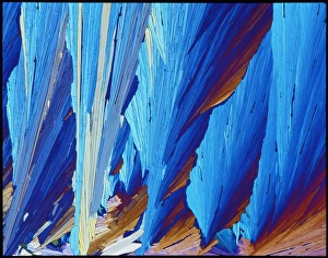
Background imageMagnified Image Collection: Silver nitrate crystals, LM

Silver nitrate crystals, LM
Silver nitrate crystals. Polarised light micrograph (LM) of silver nitrate (AgNO3) crystals. Silver nitrate is used in photography and as a test for chloride, bromide and iodide ions

Background imageMagnified Image Collection: Magnesium chloride crystals, LM

Magnesium chloride crystals, LM
Magnesium chloride. Polarised light micrograph (LM) of crystals of magnesium chloride (MgCl2). Typically, magnesium chloride forms complexes with other molecules; the hygroscopic hexahydrate

Background imageMagnified Image Collection: Magnesium sulphate crystals, LM

Magnesium sulphate crystals, LM
Magnesium sulphate. Polarised light micrograph of crystals of magnesium sulphate (MgSO4), a chemical used medicinally and in the textile industry

Background imageMagnified Image Collection: Sperm on egg during fertilisation

Sperm on egg during fertilisation
Fertilisation. Coloured scanning electron micro- graph (SEM) of sperm on the surface of a human egg (ovum) during fertilisation. The egg has a furry surface (brown)

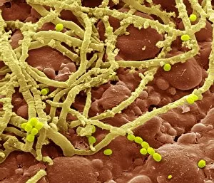
Background imageMagnified Image Collection: Bread mould (Penicillium sp. ), SEM

Bread mould (Penicillium sp. ), SEM
Bread mould. Coloured scanning electronmicrograph (SEM) of Penicillium sp. mouldgrowing on bread. The yellow fibrous structuresare hyphae, which make up the main body of thefungus

Background imageMagnified Image Collection: Proteus vulgaris bacterium, TEM

Proteus vulgaris bacterium, TEM
Proteus vulgaris bacterium, colouredtransmission electron micrograph (TEM). The yellowthreads are flagella, hair-like structures thatthe bacterium beats for motility

Background imageMagnified Image Collection: Staphylococcus aureus dividing, TEM

Staphylococcus aureus dividing, TEM
Staphylococcus aureus bacteria dividing, coloured transmission electron micrograph. Atlower left is a single bacterium, the bacterium atupper centre is starting to divide

Background imageMagnified Image Collection: Oral bacteria, TEM

Oral bacteria, TEM
Oral bacteria. Computer-enhanced colouredtransmission electron micrograph (TEM) of Porphyromonas gingivalis bacteria (formerlynamed Bacteroides gingivalis )

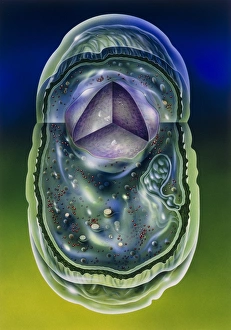
Background imageMagnified Image Collection: Bacterial cell

Bacterial cell, computer artwork. This is ageneralised bacterium, illustrating features ofdifferent bacterial types that would not becombined in a single cell

Background imageMagnified Image Collection: Helicobacter pylori bacteria in stomach

Helicobacter pylori bacteria in stomach
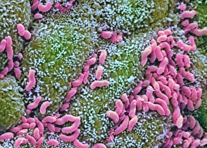
Helicobacter pylori bacteria. Colouredscanning electron micrograph (SEM) of Helicobacter pylori bacteria (pink), a causeof gastritis (stomach lining inflammation)

Background imageMagnified Image Collection: Listeria monocytogenes bacteria, TEM

Listeria monocytogenes bacteria, TEM
Listeria bacteria. Coloured transmissionelectron micrograph (TEM) of a Listeria monocytogenes bacterium (centre to bottomright). It is dividing by a process of unequalcell growth known as budding

Background imageMagnified Image Collection: Unidentified rod-shaped bacteria

Unidentified rod-shaped bacteria
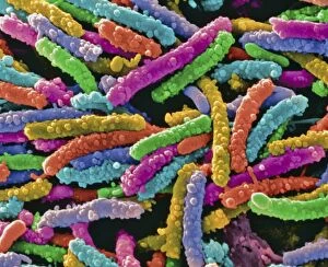
Bacteria. Coloured scanning electronmicrograph (SEM) of unidentified bacilli(rod-shaped bacteria). Magnification unknown


All Professionally Made to Order for Quick Shipping

"Exploring the unseen wonders of the microscopic world: from particle tracks to intricate tissues and beyond. " "Unveiling the hidden beauty within: a closer look at cerebellum tissue through a mesmerizing light micrograph. " "A captivating glimpse into the mysteries of subatomic particles: behold the bubble chamber photo capturing sigma particle decay. " "The groundbreaking moment in science history: witness the first observation of omega-minus particle, forever changing our understanding of matter. " "Awe-inspiring beginnings: marvel at the intricate structure of a human blastocyst, where life takes its first steps. " "Diving deep into neural networks: an illuminating immunofluorescent LM reveals stunning neurons and astrocytes in all their glory. " "Unraveling nature's cycle within us: witness the uterus lining during menstruation, as seen through SEM imaging - a fascinating display of renewal and change. " "Peering into viral threats that loom above us all: explore avian flu virus up close, revealing its complex structure and potential dangers it holds. " "The crystalline beauty behind love and bonding: discover oxytocin hormone crystals under PLM C016 / 7196 microscopy - nature's secret ingredient for connection. " "A tapestry woven by nature itself: delve into intricately detailed fabric structures captured through SEM imaging - artistry on a microscopic scale. " "Glimpsing into windows to our souls with breathtaking precision: explore the iris of an eye like never before, revealing unique patterns that make each person truly one-of-a-kind. " "Revealing fragility beneath strength: examine osteoporotic bone under SEM, shedding light on this silent disease affecting millions worldwide.