Fraud Blocker Skip to main content

Magnified Image Collection (page 7)

"Exploring the unseen wonders of the microscopic world: from particle tracks to intricate tissues and beyond

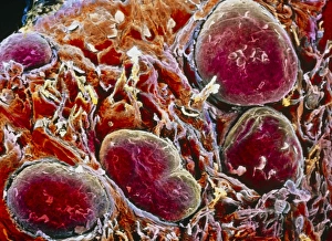
Background imageMagnified Image Collection: Renal corpuscle of kidney

Renal corpuscle of kidney
Coloured scanning electron micrograph (SEM) of a renal corpuscle in the kidney. The renal corpuscle is formed by the glomerulus (red), the convoluted capillary at centre

Background imageMagnified Image Collection: False-colour SEM of a taste bud on tongue surface

False-colour SEM of a taste bud on tongue surface
Taste bud. False-colour scanning electron micrograph of the apical part of a taste bud (pink at centre), known as a gustatory caliculus, on the dorsal surface of the tongue

Background imageMagnified Image Collection: False-colour SEM of fungiform papillae on tongue

False-colour SEM of fungiform papillae on tongue

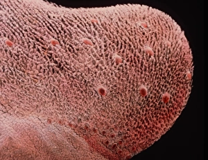
Background imageMagnified Image Collection: False-colour SEM of dorsal surface of tongue

False-colour SEM of dorsal surface of tongue
Tongue surface. False-colour scanning electron micrograph (SEM) of the dorsal surface of the tongue. It is covered by two types of projections known as filiform and fungiform papillae

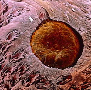
Background imageMagnified Image Collection: False-colour SEM of a papilla vallata

False-colour SEM of a papilla vallata
Papilla vallata on tongues surface. False-colour scanning electron micrograph of a papilla vallata (brown), a site of taste buds, partly surrounded by filiform papillae (bottom)

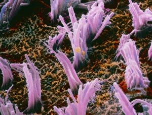
Background imageMagnified Image Collection: False-colour SEM of ciliated hair cells in the ear

False-colour SEM of ciliated hair cells in the ear
Inner ear. False-colour scanning electron micrograph of bundles of ciliated hair cells (pink) situated in the macula utriculi within the human inner ear

Background imageMagnified Image Collection: False-colour SEM of hair cells in the inner ear

False-colour SEM of hair cells in the inner ear
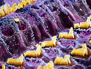
Inner ear. False-colour scanning electron micrograph of hair cells (yellow) which are part of the Organ of Corti in the inner ear

Background imageMagnified Image Collection: Coloured SEM of iris epithelial cells of the eye

Coloured SEM of iris epithelial cells of the eye
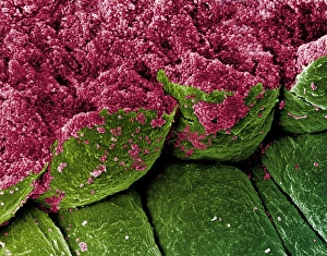
Iris epithelium. Coloured scanning electron micrograph (SEM) of epithelial cells (green) lining the underside of the human iris

Background imageMagnified Image Collection: Blood vessels in eye

Blood vessels in eye
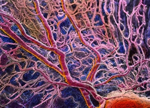
False-colour scanning electron micrograph (SEM) of blood vessels in the choroid of the eye. A branch- ing network of arteries and veins can be seen in this area under the central fovea

Background imageMagnified Image Collection: Light micrograph of cardiac muscle & fatty disease

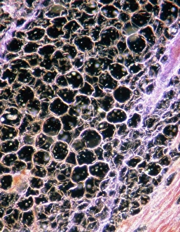
Light micrograph of cardiac muscle & fatty disease

Background imageMagnified Image Collection: False-colour SEM of cells from lens of hu

False-colour SEM of cells from lens of hu

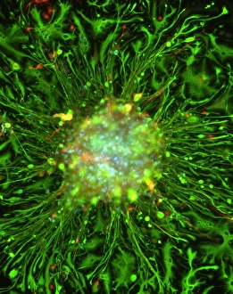
Background imageMagnified Image Collection: Neurosphere culture

Neurosphere culture. Fluorescent light microscope of a group of neural stem cells (neurosphere) in culture. The stem cells are differentiating into neurons (red) and nerve support cells (green)

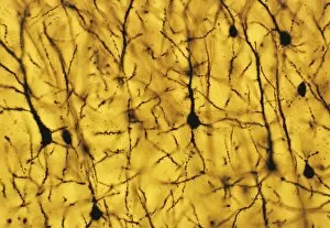
Background imageMagnified Image Collection: Cerebral cortex nerve cells

Cerebral cortex nerve cells

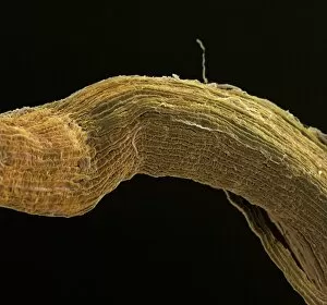
Background imageMagnified Image Collection: Nerve fibre, SEM

Nerve fibre, SEM
Nerve fibres. Coloured scanning electron micrograph (SEM) of nerve fibres (brown). A group of nerves such as this is known as a fasciculus

Background imageMagnified Image Collection: Blood clot crystals, SEM

Blood clot crystals, SEM
Blood clot crystals. Coloured scanning electron micrograph (SEM) of crystals of albumin from a blood clot. Albumin is the most abundant protein in the blood plasma

Background imageMagnified Image Collection: Coloured SEM of red blood cells forming a clot

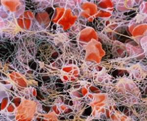
Coloured SEM of red blood cells forming a clot
Blood clot. Coloured Scanning Electron Micrograph (SEM) showing the formation of a blood clot. Red blood cells (erythrocytes)

Background imageMagnified Image Collection: Blood clot

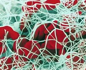
Blood clot
False-colour scanning electron micrograph (SEM) of a blood clot. Red blood cells have been trapped by a web of thin yellow-white strands of fibrin

Background imageMagnified Image Collection: Red blood cell crenation, SEM

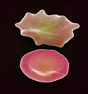
Red blood cell crenation, SEM
Red blood cell crenation. Coloured scanning electron micrograph (SEM) of two red blood cells (erythrocytes). The cell on the bottom is normal

Background imageMagnified Image Collection: Red blood cells, SEM

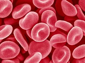
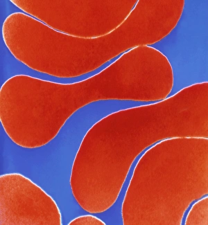
Red blood cells, SEM
Red blood cells. Coloured scanning electron micrograph (SEM) of red blood cells (erythrocytes). Red blood cells are biconcave, disc-shaped cells that transport oxygen from the lungs to body cells

Background imageMagnified Image Collection: Colour TEM of red blood cells, rouleau formation

Colour TEM of red blood cells, rouleau formation
Red blood cells. Coloured Transmission Electron Micrograph (TEM) of sectioned human red blood cells (erythrocytes) in a rouleau formation

Background imageMagnified Image Collection: Coloured SEM of red blood cells in blood vessel

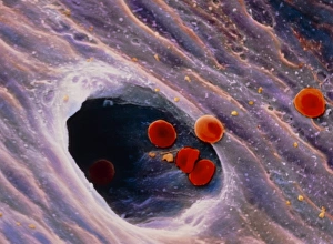
Coloured SEM of red blood cells in blood vessel
Red blood cells. Coloured scanning electron micrograph of a group of red blood cells (erythrocytes). They are travelling through a large vessel and are just entering a small vessel

Background imageMagnified Image Collection: SEM of bone marrow

SEM of bone marrow

Background imageMagnified Image Collection: Tendon fibres, TEM

Tendon fibres, TEM
Tendon fibres, coloured transmission electron micrograph (TEM). A tendon is made up of parallel bundles of collagen fibres

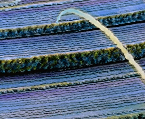
Background imageMagnified Image Collection: Coloured SEM of skeletal (striated) muscle fibres

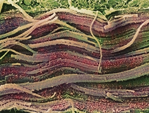
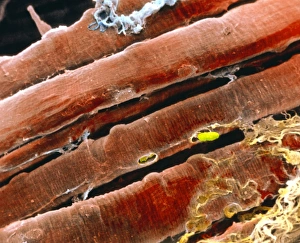
Coloured SEM of skeletal (striated) muscle fibres
Skeletal muscle. Coloured scanning electron micrograph (SEM) of skeletal muscle fibres. Muscle fibres are extremely long, cylindrical cells

Background imageMagnified Image Collection: Coloured SEM of a bundle of skeletal muscle fibres

Coloured SEM of a bundle of skeletal muscle fibres

Background imageMagnified Image Collection: False-colour SEM of skeletal muscle fibres

False-colour SEM of skeletal muscle fibres
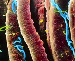
Muscle. False-colour scanning electron micrograph of a bundle of skeletal muscle fibres (red), also known as striated muscle fibres, and a few capillaries (blue)

Background imageMagnified Image Collection: Fossilised compact bone, SEM

Fossilised compact bone, SEM
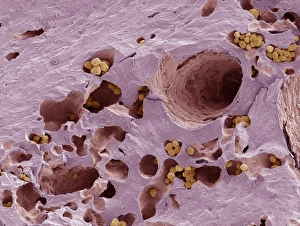
Fossilised compact bone. Coloured scanning electron micrograph (SEM) of a section through fossilised compact bone. This tissue is found in the dense walls of the shafts of bones

Background imageMagnified Image Collection: LM of cervical smear cells with mild dyskaryosis

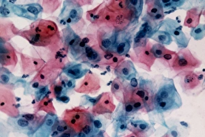
LM of cervical smear cells with mild dyskaryosis

Background imageMagnified Image Collection: Vaccinia virus particles, SEM

Vaccinia virus particles, SEM
Vaccinia virus particles. Coloured scanning electron micrograph (SEM) of vaccinia virus particles (red) budding from a cell (blue and green) after replicating

Background imageMagnified Image Collection: HIV particles, computer artwork

HIV particles, computer artwork. HIV (human immunodeficiency virus) causes AIDS (acquired immune deficiency syndrome). The virus consists of an RNA genome

Background imageMagnified Image Collection: West Nile viruses

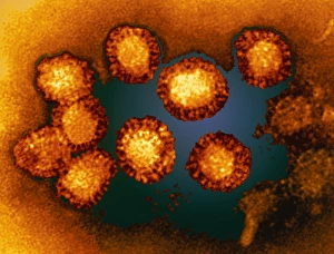
West Nile viruses. Coloured transmission electron micrograph (TEM) of a group of West Nile viruses (WNV). This virus is known to cause encephalitis in humans

Background imageMagnified Image Collection: Virus and red blood cells

Virus and red blood cells. Computer artwork of a virus under a magnifying glass surrounded by red blood cells. Viruses consist of a small core of genetic material


All Professionally Made to Order for Quick Shipping

"Exploring the unseen wonders of the microscopic world: from particle tracks to intricate tissues and beyond. " "Unveiling the hidden beauty within: a closer look at cerebellum tissue through a mesmerizing light micrograph. " "A captivating glimpse into the mysteries of subatomic particles: behold the bubble chamber photo capturing sigma particle decay. " "The groundbreaking moment in science history: witness the first observation of omega-minus particle, forever changing our understanding of matter. " "Awe-inspiring beginnings: marvel at the intricate structure of a human blastocyst, where life takes its first steps. " "Diving deep into neural networks: an illuminating immunofluorescent LM reveals stunning neurons and astrocytes in all their glory. " "Unraveling nature's cycle within us: witness the uterus lining during menstruation, as seen through SEM imaging - a fascinating display of renewal and change. " "Peering into viral threats that loom above us all: explore avian flu virus up close, revealing its complex structure and potential dangers it holds. " "The crystalline beauty behind love and bonding: discover oxytocin hormone crystals under PLM C016 / 7196 microscopy - nature's secret ingredient for connection. " "A tapestry woven by nature itself: delve into intricately detailed fabric structures captured through SEM imaging - artistry on a microscopic scale. " "Glimpsing into windows to our souls with breathtaking precision: explore the iris of an eye like never before, revealing unique patterns that make each person truly one-of-a-kind. " "Revealing fragility beneath strength: examine osteoporotic bone under SEM, shedding light on this silent disease affecting millions worldwide.