Fraud Blocker Skip to main content

Human Anatomy Collection (page 3)

"Exploring the Intricacies of Human Anatomy: From Knees to Brains and Everything In Between" Normal knees

Background imageHuman Anatomy Collection: Albinus I, Pl. IX: Musculature, illustration from Tabulae sceleti et musculorum

Albinus I, Pl. IX: Musculature, illustration from Tabulae sceleti et musculorum
HMB306108 Albinus I, Pl. IX: Musculature, illustration from Tabulae sceleti et musculorum corporis humani, by Bernhard Siegfried Albinus (1697-1770), published by J.&H. Verbeek, bibliop

Background imageHuman Anatomy Collection: anatomical view of a tooth, the source of toothache is shown as a demon of hell

anatomical view of a tooth, the source of toothache is shown as a demon of hell
5892858 anatomical view of a tooth, the source of toothache is shown as a demon of hell (watercolour and gold powder on paper) by Turkish School

Background imageHuman Anatomy Collection: Albinus I, Pl. I: Skeleton, illustration from Tabulae sceleti et musculorum corporis

Albinus I, Pl. I: Skeleton, illustration from Tabulae sceleti et musculorum corporis
HMB306109 Albinus I, Pl. I: Skeleton, illustration from Tabulae sceleti et musculorum corporis humani, by Bernhard Siegfried Albinus (1697-1770), published by J.&H. Verbeek, bibliop

Background imageHuman Anatomy Collection: Albinus I, Pl. III: Skeleton, illustration from Tabulae sceleti et musculorum

Albinus I, Pl. III: Skeleton, illustration from Tabulae sceleti et musculorum
HMB306105 Albinus I, Pl. III: Skeleton, illustration from Tabulae sceleti et musculorum corporis humani, by Bernhard Siegfried Albinus (1697-1770), published by J.&H. Verbeek, bibliop

Background imageHuman Anatomy Collection: Albinus II, Pl. X: Cervical vertebra, illustration from Tabulae ossium humanorum

Albinus II, Pl. X: Cervical vertebra, illustration from Tabulae ossium humanorum
HMB306104 Albinus II, Pl. X: Cervical vertebra, illustration from Tabulae ossium humanorum, by Bernhard Siegfried Albinus (1697-1770), published by J.&H. Verbeek, bibliop

Background imageHuman Anatomy Collection: Albinus I, Pl. II: Skeleton, illustration from Tabulae sceleti et musculorum

Albinus I, Pl. II: Skeleton, illustration from Tabulae sceleti et musculorum
HMB306107 Albinus I, Pl. II: Skeleton, illustration from Tabulae sceleti et musculorum corporis humani, by Bernhard Siegfried Albinus (1697-1770), published by J.&H. Verbeek, bibliop

Background imageHuman Anatomy Collection: Albinus I, Dedication page, from Tabulae sceleti et musculorum corporis humani

Albinus I, Dedication page, from Tabulae sceleti et musculorum corporis humani
HMB306113 Albinus I, Dedication page, from Tabulae sceleti et musculorum corporis humani, by Bernhard Siegfried Albinus (1697-1770), published by J.&H. Verbeek, bibliop

Background imageHuman Anatomy Collection: Albinus II, Pl. II, Skull, illustration from Tabulae ossium humanorum

Albinus II, Pl. II, Skull, illustration from Tabulae ossium humanorum
HMB306102 Albinus II, Pl. II, Skull, illustration from Tabulae ossium humanorum, by Bernhard Siegfried Albinus (1697-1770), published by J.&H. Verbeek, bibliop

Background imageHuman Anatomy Collection: Tom Jones, singer from Wales, pictured circa 1965, examining his famous tonsils in

Tom Jones, singer from Wales, pictured circa 1965, examining his famous tonsils in the mirror. Sir Thomas John Woodward OBE (born 7 June 1940), known professionally as Tom Jones, is a Welsh singer

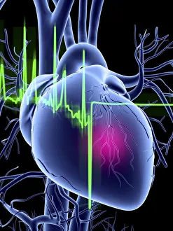
Background imageHuman Anatomy Collection: Heart attack and ECG trace

Heart attack and ECG trace
Heart attack, conceptual computer artwork showing an ECG (electrocardiogram) trace from an unhealthy human heart. An ECG uses electrodes to measure the electrical activity of the heart during

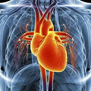
Background imageHuman Anatomy Collection: Chest anatomy, artwork

Chest anatomy, artwork
Chest anatomy, computer artwork

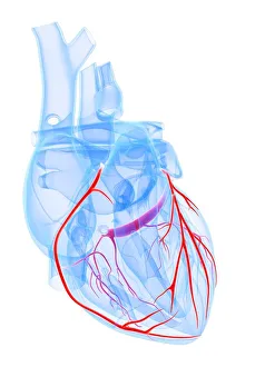
Background imageHuman Anatomy Collection: Healthy heart, artwork

Healthy heart, artwork
Healthy heart. Computer artwork showing the coronary arteries

Background imageHuman Anatomy Collection: Aortic aneurysm, illustration

Aortic aneurysm, illustration
Aortic aneurysm. Computer illustration showing an aneurysm in the ascending aorta

Background imageHuman Anatomy Collection: Angioplasty, artwork

Angioplasty, artwork
Balloon angioplasty. Computer artwork of a stent being placed in a narrowed blood vessel

Background imageHuman Anatomy Collection: Cardiovascular system, artwork

Cardiovascular system, artwork
Cardiovascular system, computer artwork

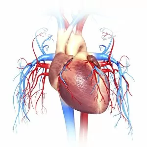
Background imageHuman Anatomy Collection: Male heart, illustration

Male heart, illustration
Illustration of male heart and arteries

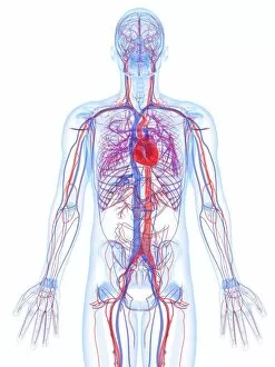
Background imageHuman Anatomy Collection: Male cardiovascular system, artwork

Male cardiovascular system, artwork
Male cardiovascular system, computer artwork

Background imageHuman Anatomy Collection: Human heart attack, illustration

Human heart attack, illustration
Human heart attack, computer illustration

Background imageHuman Anatomy Collection: Human vascular system, artwork

Human vascular system, artwork
Human vascular system, computer artwork

Background imageHuman Anatomy Collection: Human heart, artwork

Human heart, artwork
Human heart, computer artwork

Background imageHuman Anatomy Collection: Human heart, computer artwork

Human heart, computer artwork

Background imageHuman Anatomy Collection: A leg and knee injury sustained from a repetitive motion

A leg and knee injury sustained from a repetitive motion
Fitness, Sport, Vest, Anatomy, Diagram, Muscle, Illustration, Bone, Repetition, Athlete, Shorts, Leg, Injury, Medical, Jumping, Biology, Knee, See Through, Weekend Activities, Hurdle

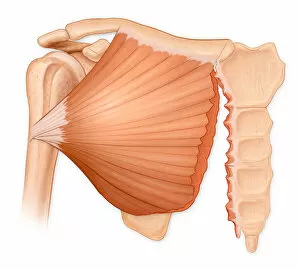
Background imageHuman Anatomy Collection: Microscopic muscle fiber tears of the pectoralis major muscle

Microscopic muscle fiber tears of the pectoralis major muscle
Artwork, Bone, Clavicle, Human Anatomy, humerus, Illustration, inflammation, Injury, Medical, Microscopic, Muscle, muscle fiber tear, Pectoralis major muscle, Pectoralis Major Tear Syndrome, scapula

Background imageHuman Anatomy Collection: Complete rupture of the pectoralis major tendon from the humerus

Complete rupture of the pectoralis major tendon from the humerus
Horizontal, Detail, Anatomy, Diagram, Muscle, Illustration, Bone, Injury, Medical, Biology, Bicep, Cut Out, White Background, Artwork, Color Image, Close Up, Healthcare And Medicine, The Human Body

Background imageHuman Anatomy Collection: Pectoralis muscle tear, associated with a hematoma formation and cosmetic deformity

Pectoralis muscle tear, associated with a hematoma formation and cosmetic deformity
Horizontal, Detail, Anatomy, Diagram, Muscle, Illustration, Bone, Injury, Medical, Biology, Cut Out, White Background, Artwork, Color Image, Close Up, Healthcare And Medicine, The Human Body

Background imageHuman Anatomy Collection: Normal posterior view of the hip bones and gluteus maximus muscle including the lumbar

Normal posterior view of the hip bones and gluteus maximus muscle including the lumbar
Detail, Anatomy, Diagram, Muscle, Illustration, Bone, Medical, Hip, Biology, Pelvis, Femur, See Through, Rear View, Cut Out, White Background, Artwork, Color Image, Close Up, Healthcare And Medicine

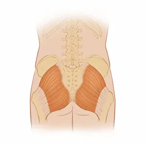
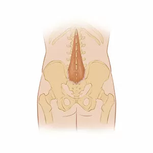
Background imageHuman Anatomy Collection: Normal posterior view of the back highlighting the multifidus muscle

Normal posterior view of the back highlighting the multifidus muscle
Detail, Anatomy, Diagram, Muscle, Illustration, Ribs, Bone, Medical, Biology, Pelvis, See Through, Rear View, Cut Out, White Background, Artwork, Color Image, Close Up, Healthcare And Medicine

Background imageHuman Anatomy Collection: Back pain, conceptual artwork

Back pain, conceptual artwork
Back pain, conceptual computer artwork

Background imageHuman Anatomy Collection: Healthy small intestines, artwork

Healthy small intestines, artwork
Healthy small intestines, computer artwork

Background imageHuman Anatomy Collection: Upper body bones, artwork

Upper body bones, artwork
Upper body bones, computer artwork

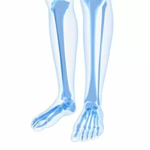
Background imageHuman Anatomy Collection: Leg bones, artwork

Leg bones, artwork
Leg bones, computer artwork

Background imageHuman Anatomy Collection: Normal childs head, X-ray

Normal childs head, X-ray
Normal childs head. Coloured X-ray of the head of a 13 year old

Background imageHuman Anatomy Collection: Illustration of human arm bones

Illustration of human arm bones
Medicine: Human anatomy, bones, arm. Drawing

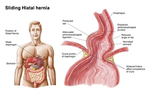
Background imageHuman Anatomy Collection: Medical ilustration of a hiatal hernia in the upper part of the stomach into the thorax

Medical ilustration of a hiatal hernia in the upper part of the stomach into the thorax

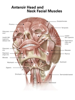
Background imageHuman Anatomy Collection: Anterior neck and facial muscles of the human head (with labels)

Anterior neck and facial muscles of the human head (with labels)

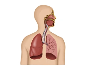
Background imageHuman Anatomy Collection: Anatomy of human respiratory system

Anatomy of human respiratory system

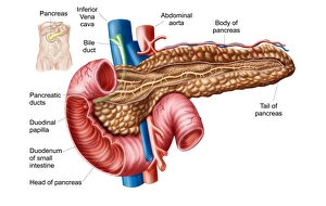
Background imageHuman Anatomy Collection: Anatomy of pancreas

Anatomy of pancreas

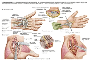
Background imageHuman Anatomy Collection: Medical illustration showing carpal tunnel syndrome in the human wrist

Medical illustration showing carpal tunnel syndrome in the human wrist, and the surgical procedures associated with it

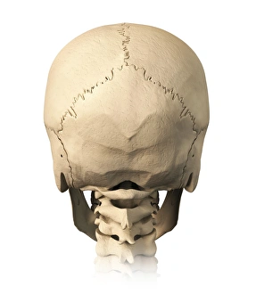
Background imageHuman Anatomy Collection: Anatomy of human skull, rear view

Anatomy of human skull, rear view

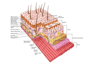
Background imageHuman Anatomy Collection: Anatomy of the human skin

Anatomy of the human skin

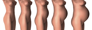
Background imageHuman Anatomy Collection: Growth of female midsection during pregnancy stages

Growth of female midsection during pregnancy stages

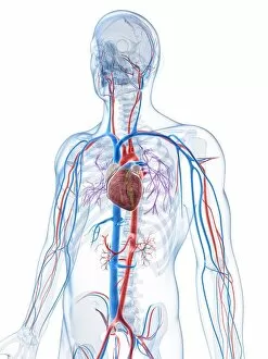
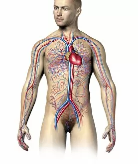
Background imageHuman Anatomy Collection: Male human circulatory system, upper body

Male human circulatory system, upper body
Male human circulatory system, upper part, superimposed to realistic body

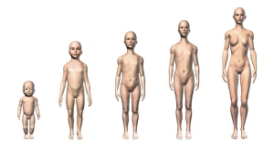
Background imageHuman Anatomy Collection: Female human body scheme of different age stages

Female human body scheme of different age stages, showing five different ages with relative body shapes

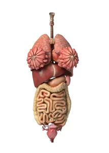
Background imageHuman Anatomy Collection: 3D rendering of healthy female internal organs

3D rendering of healthy female internal organs

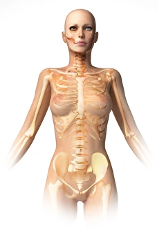
Background imageHuman Anatomy Collection: Female body with bone skeleton and internal organs superimposed

Female body with bone skeleton and internal organs superimposed
Anatomy of female body with bone skeleton and all internal organs superimposed

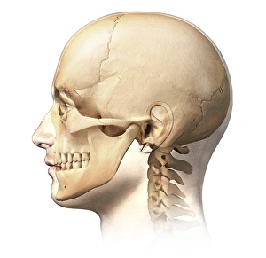
Background imageHuman Anatomy Collection: Male human head with skull in ghost effect, side view

Male human head with skull in ghost effect, side view. on white background

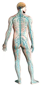
Background imageHuman Anatomy Collection: Diagram of human nervous system, posterior view

Diagram of human nervous system, posterior view


All Professionally Made to Order for Quick Shipping

"Exploring the Intricacies of Human Anatomy: From Knees to Brains and Everything In Between" Normal knees, X-ray: A glimpse into the intricate structure of our knee joints, highlighting their normalcy. Anatomy of human brain, inferior view: Unveiling the hidden complexities within our brains from a unique perspective. Facial muscles of the human face (with labels): Discovering the intricate network of muscles that allow us to express emotions and communicate non-verbally. Anatomy of human knee joint: Delving deep into the mechanics behind one of our most crucial joints, revealing its inner workings. Superior view of human brain with colored lobes and labels: An illuminating look at our brain's superior side, showcasing distinct lobes responsible for various functions. The human body with superimposed colored plates by Julien Bougle: Witnessing an artistic representation where vibrant colors highlight different systems within our bodies in a visually captivating manner. Cross-section diagram of human mouth and jaw: Peering inside our oral cavity to understand how teeth, gums, and jaws work together seamlessly for speech and mastication. Human foot anatomy showing skin, veins, arteries, muscles, and bones: Uncovering the complexity beneath every step we take – from delicate skin to robust bones – as we explore foot anatomy in detail. Healthy knee, X-ray: Appreciating a radiographic image that showcases a healthy knee joint functioning optimally without any abnormalities or injuries. Illustration showing intestinal villi: Journeying through microscopic structures called villi lining our intestines that aid in nutrient absorption - an essential aspect often overlooked but vital for digestion.