Fraud Blocker Skip to main content

Human Anatomy Collection (page 27)

"Exploring the Intricacies of Human Anatomy: From Knees to Brains and Everything In Between" Normal knees

Background imageHuman Anatomy Collection: Thomas Willis (1621-1672), physician and anatomist, XIX century, engraving

Thomas Willis (1621-1672), physician and anatomist, XIX century, engraving
Physicians and scholars: Great Britain - XVII century. Thomas Willis (1621-1672), physician and anatomist. XIX century, engraving

Background imageHuman Anatomy Collection: Gaspare Aselli (1581-1626), physician, 19th century, engraving

Gaspare Aselli (1581-1626), physician, 19th century, engraving
Italy - 16th-17th century. Gaspare Aselli (1581-1626), physician. 19th century, engraving

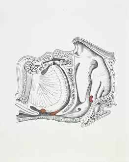
Background imageHuman Anatomy Collection: Illustration of tonsils

Illustration of tonsils
Medicine: tonsils. Drawing

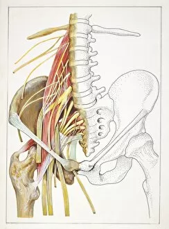
Background imageHuman Anatomy Collection: Illustration of lumbago, lumbar and pelvic regions

Illustration of lumbago, lumbar and pelvic regions
Medicine: lumbago, lumbar and pelvic regions. Drawing

Background imageHuman Anatomy Collection: Giambattista Morgagni (1682-1771), physician, engraving

Giambattista Morgagni (1682-1771), physician, engraving
Physicians and scholars: Italy - 18th century. Giambattista Morgagni (1682-1771), physician. Engraving

Background imageHuman Anatomy Collection: Giorgio Baglivi (1668-1707), anatomist and pathologist, 18th century, engraving

Giorgio Baglivi (1668-1707), anatomist and pathologist, 18th century, engraving
Physicians and scholars: Italy - 17th-18th century. Giorgio Baglivi (1668-1707), anatomist and pathologist. 18th century, engraving

Background imageHuman Anatomy Collection: Giovanni Battista Canano (1515-1579), anatomist, 19th century, engraving

Giovanni Battista Canano (1515-1579), anatomist, 19th century, engraving
Physicians and scholars: Italy - 16th century. Giovanni Battista Canano (1515-1579), anatomist. 19th century, engraving

Background imageHuman Anatomy Collection: Guy de Chauliac (1300-1368), physician, 18th century, engraving

Guy de Chauliac (1300-1368), physician, 18th century, engraving
Physicians and scholars: France - 16th century. Guy de Chauliac (1300-1368), physician. 18th century, engraving

Background imageHuman Anatomy Collection: Giambattista Morgagni (1682-1771), physician and anatomist, engraving

Giambattista Morgagni (1682-1771), physician and anatomist, engraving
Italy - 18th century. Giambattista Morgagni (1682-1771), physician and anatomist. Engraving

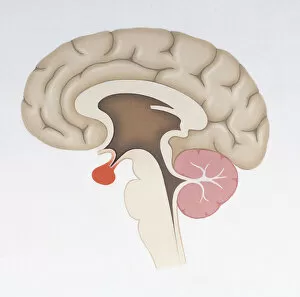
Background imageHuman Anatomy Collection: Human brain

Human brain

Background imageHuman Anatomy Collection: Tongue, and various papilla areas; vallate, fungiform and filiform

Tongue, and various papilla areas; vallate, fungiform and filiform. Also shows the four taste areas of sweet, salt, sour and bitter

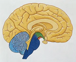
Background imageHuman Anatomy Collection: Basic cutaway illustration of the human brain showing the thalamus, cerebrum, hypothalmus

Basic cutaway illustration of the human brain showing the thalamus, cerebrum, hypothalmus, cerebellum and brain stem

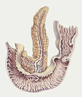
Background imageHuman Anatomy Collection: Medicine: Human body, Duodenum and pancreas, illustration

Medicine: Human body, Duodenum and pancreas, illustration Medicine: Human anatomy

Background imageHuman Anatomy Collection: Zoology: Birds - Passeriformes - Western Orphean Warbler (Sylvia hortensis). Art work

Zoology: Birds - Passeriformes - Western Orphean Warbler (Sylvia hortensis). Art work


All Professionally Made to Order for Quick Shipping

"Exploring the Intricacies of Human Anatomy: From Knees to Brains and Everything In Between" Normal knees, X-ray: A glimpse into the intricate structure of our knee joints, highlighting their normalcy. Anatomy of human brain, inferior view: Unveiling the hidden complexities within our brains from a unique perspective. Facial muscles of the human face (with labels): Discovering the intricate network of muscles that allow us to express emotions and communicate non-verbally. Anatomy of human knee joint: Delving deep into the mechanics behind one of our most crucial joints, revealing its inner workings. Superior view of human brain with colored lobes and labels: An illuminating look at our brain's superior side, showcasing distinct lobes responsible for various functions. The human body with superimposed colored plates by Julien Bougle: Witnessing an artistic representation where vibrant colors highlight different systems within our bodies in a visually captivating manner. Cross-section diagram of human mouth and jaw: Peering inside our oral cavity to understand how teeth, gums, and jaws work together seamlessly for speech and mastication. Human foot anatomy showing skin, veins, arteries, muscles, and bones: Uncovering the complexity beneath every step we take – from delicate skin to robust bones – as we explore foot anatomy in detail. Healthy knee, X-ray: Appreciating a radiographic image that showcases a healthy knee joint functioning optimally without any abnormalities or injuries. Illustration showing intestinal villi: Journeying through microscopic structures called villi lining our intestines that aid in nutrient absorption - an essential aspect often overlooked but vital for digestion.