
Human Anatomy Collection (page 2)
"Exploring the Intricacies of Human Anatomy: From Knees to Brains and Everything In Between" Normal knees


All Professionally Made to Order for Quick Shipping
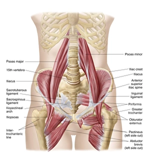
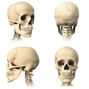
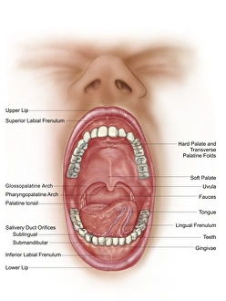
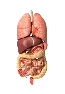
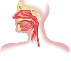
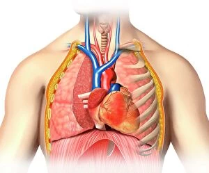
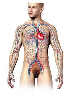
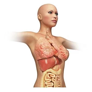
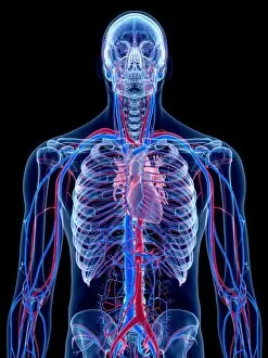
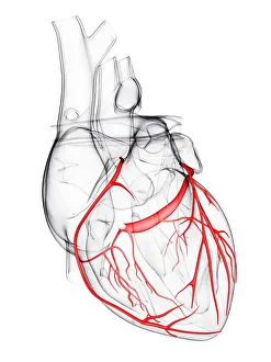
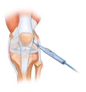
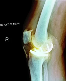
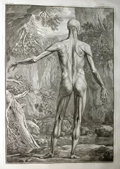
"Exploring the Intricacies of Human Anatomy: From Knees to Brains and Everything In Between" Normal knees, X-ray: A glimpse into the intricate structure of our knee joints, highlighting their normalcy. Anatomy of human brain, inferior view: Unveiling the hidden complexities within our brains from a unique perspective. Facial muscles of the human face (with labels): Discovering the intricate network of muscles that allow us to express emotions and communicate non-verbally. Anatomy of human knee joint: Delving deep into the mechanics behind one of our most crucial joints, revealing its inner workings. Superior view of human brain with colored lobes and labels: An illuminating look at our brain's superior side, showcasing distinct lobes responsible for various functions. The human body with superimposed colored plates by Julien Bougle: Witnessing an artistic representation where vibrant colors highlight different systems within our bodies in a visually captivating manner. Cross-section diagram of human mouth and jaw: Peering inside our oral cavity to understand how teeth, gums, and jaws work together seamlessly for speech and mastication. Human foot anatomy showing skin, veins, arteries, muscles, and bones: Uncovering the complexity beneath every step we take – from delicate skin to robust bones – as we explore foot anatomy in detail. Healthy knee, X-ray: Appreciating a radiographic image that showcases a healthy knee joint functioning optimally without any abnormalities or injuries. Illustration showing intestinal villi: Journeying through microscopic structures called villi lining our intestines that aid in nutrient absorption - an essential aspect often overlooked but vital for digestion.