Fraud Blocker Skip to main content

Fractured Collection (page 4)

"Fractured: A Visual Journey of Broken Bones and Historical Moments" The X-ray reveals a broken wrist bone, a painful reminder of an unfortunate accident

Background imageFractured Collection: Blitz in London -- Standish Road, Hammersmith, WW2

Blitz in London -- Standish Road, Hammersmith, WW2
Blitz in London -- repairing a fractured water main in Standish Road, King Street, Hammersmith, 4 September 1940. A bomb-damaged house can be seen, top left

Background imageFractured Collection: Fractured pollen grain

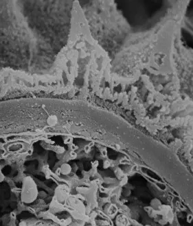
Fractured pollen grain
Scanning electron microscope (SEM) image showing a fractured pollen grain

Background imageFractured Collection: Fractured anther

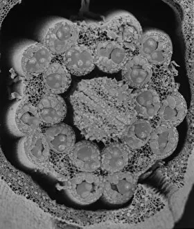
Fractured anther
Scanning electron microscope (SEM) image showing a fractured anther, otherwise known as the sac, which contains the pollen in the male sex organs (stamens)

Background imageFractured Collection: Fractured barrel of German Paris Gun, WW1

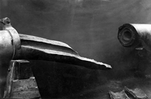
Fractured barrel of German Paris Gun, WW1
The fractured barrel of the German Paris Gun (also known as the Emperor William Gun), a long-range seige gun designed and manufactured by Krupp, used towards the end of the First World War

Background imageFractured Collection: Dog with Jaw Splint

Dog with Jaw Splint
A fixation splint for a fractured jaw, the latest development. Date: circa 1960

Background imageFractured Collection: Swallow, X-ray

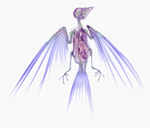
Swallow, X-ray
Swallow. Coloured X-ray of a swallow (family Hirundidae). The swallow spends much of its time on the wing, and it is well adapted for energy- efficient flight; its hollow bones

Background imageFractured Collection: Hair, SEM

Hair, SEM
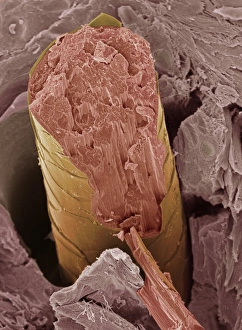
Hair fracture. Coloured scanning electron micrograph (SEM) of a fractured hair (yellow and pink). The surrounding layer of facial skin (brown) has a typically scale-like surface

Background imageFractured Collection: Cats tooth, SEM

Cats tooth, SEM
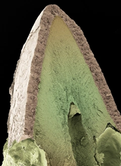
Cats tooth. Coloured scanning electron micrograph (SEM) of a vertical fracture through a canine tooth of a domestic cat (Felis catus)

Background imageFractured Collection: Small intestine villus, SEM

Small intestine villus, SEM
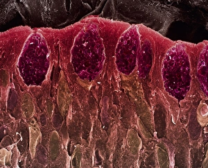
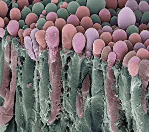
Small intestine villus. Coloured scanning electron micrograph (SEM) of a freeze fracture section through a villus from the mucosal lining of the small intestine

Background imageFractured Collection: Secretory cells in pancreas, SEM

Secretory cells in pancreas, SEM
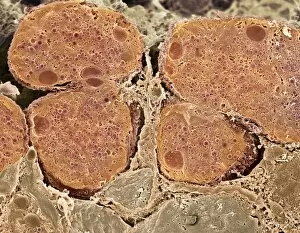
Pancreatic secretory cells. Coloured scanning electron micrograph (SEM) of a freeze-fracture through a healthy pancreas, showing the secretory tissue

Background imageFractured Collection: Pancreas cells, SEM

Pancreas cells, SEM
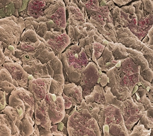
Pancreas cells. Coloured scanning electron micrograph (SEM) of acinar (exocrine) pancreatic cells. Acinar cells produce and excrete digestive enzymes to the small intestine, via the pancreatic ducts

Background imageFractured Collection: Choroid plexus secretory cells, SEM

Choroid plexus secretory cells, SEM
Choroid plexus secretory cells. Coloured scanning electron micrograph (SEM) of a freeze-fractured choroid plexus from a brain

Background imageFractured Collection: Retina, SEM

Retina, SEM
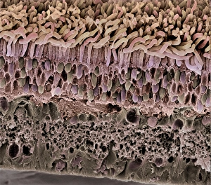
Retina. Coloured scanning electron micrograph (SEM) of a section through a freeze-fractured retina, showing the light-sensitive rods and cones and their associated neurones

Background imageFractured Collection: Compact bone, SEM

Compact bone, SEM
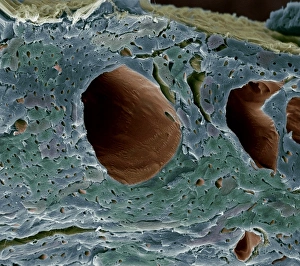
Compact bone. Coloured scanning electron micrograph (SEM) of a freeze-fractured sample of compact bone from a knee joint. This type of bone is found in the walls of bones

Background imageFractured Collection: Pinned broken jaw, X-ray

Pinned broken jaw, X-ray
Pinned broken jaw. Coloured X-ray of the skull (front view) of a 65 year old man, showing a pinned fracture of the lower jawbone (mandible)

Background imageFractured Collection: Fractured wrist, X-ray

Fractured wrist, X-ray
Broken wrist. Coloured X-ray of the hand of a patient with a fractured scaphoid bone (orange) that has failed to heal. The scaphoid bone is one of the carpel bones

Background imageFractured Collection: Broken arm in plaster cast, X-ray

Broken arm in plaster cast, X-ray
Broken arm in plaster cast. Coloured X-ray of the arm of a young woman set in a plaster cast from wrist to upper arm. The fractured bone itself has healed and so it is not clearly visible here

Background imageFractured Collection: Kneecap fracture, X-ray

Kneecap fracture, X-ray
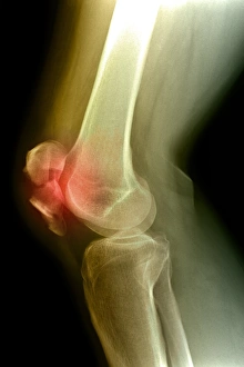
Kneecap fracture. Coloured X-ray of a broken kneecap (patella), as viewed from the side with the knee bent. The kneecap (far left, centre) is a disc of bone that covers the front of the knee joint

Background imageFractured Collection: Pinned ankle fracture, coloured X-ray

Pinned ankle fracture, coloured X-ray
Pinned ankle fracture. Coloured X-ray of the ankle bones of a patient with a pinned ankle fracture. The malleolus (bony projection at the end of a bone) of the fibula (right)

Background imageFractured Collection: Crushed broken foot, X-ray

Crushed broken foot, X-ray
Crushed broken foot. Coloured X-ray of the foot of an adult woman showing fractures to the cuneiform bones and ends of metatarsal bones of the foot. The affected area is at centre (blue)

Background imageFractured Collection: Fractured kneecap, X-ray

Fractured kneecap, X-ray
Fractured kneecap. Coloured X-ray of a side-view of the leg of a patient with a fractured kneecap (centre right). The femur (thigh bone) is at top

Background imageFractured Collection: Coloured X-ray of a fractured tibia & fibula

Coloured X-ray of a fractured tibia & fibula
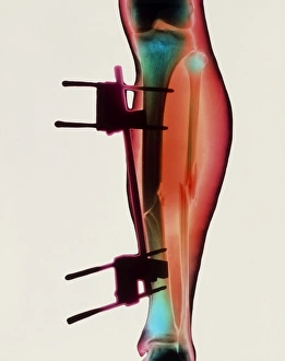
Pinned broken leg. Coloured X-ray of a pinned fracture of both the tibia and fibula (lower leg bones). The tibia (larger bone, centre) has been immobilised by metal pins (lower & upper centre)

Background imageFractured Collection: Lacrimal gland, SEM

Lacrimal gland, SEM

Background imageFractured Collection: Pinned broken leg

Pinned broken leg. X-ray of a pinned fracture of both the tibia and fibula (lower leg bones). The tibia (larger bone, at centre) has been immobilised by metal pins (lower & upper centre)

Background imageFractured Collection: Heart blood vessels, SEM

Heart blood vessels, SEM
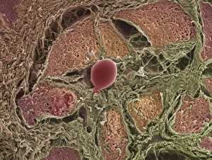
Heart blood vessels. Coloured scanning electron micrograph (SEM) of a freeze-fractured section through cardiac muscle, exposing the blood vessels of the heart

Background imageFractured Collection: Brain artery, SEM

Brain artery, SEM
Brain artery. Coloured scanning electron micrograph (SEM) of a freeze-fractured artery from the brain. The fracture plane has passed through the artery

Background imageFractured Collection: Interior of a bone, SEM

Interior of a bone, SEM
Interior of a bone. Coloured scanning electron micrograph (SEM) of a long bone fractured to show its interior. The outer layer (pink) of the bone comprises proliferating cartilage

Background imageFractured Collection: Lower leg fracture

Lower leg fracture
MODEL RELEASED. Lower leg fracture. Doctor showing a patient an x-ray of his fractured lower leg

Background imageFractured Collection: Kneecap fractures, X-ray

Kneecap fractures, X-ray
Kneecap fractures. Coloured X-ray of the kneecap (patella bone) of a 35 year old man, as viewed from above when the knee is bent, showing hairline fractures

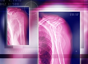
Background imageFractured Collection: Fractured shoulder, X-rays

Fractured shoulder, X-rays
Shoulder fracture, coloured X-rays. The joint is seen from the side at left and from the front at right. The joint has not been dislocated by the fracture

Background imageFractured Collection: Broken collar bone, X-ray

Broken collar bone, X-ray
Broken collar bone. Coloured frontal X-ray of a patient with a fractured left collar bone (clavicle). The fracture is at the top right of the shoulder

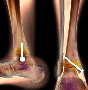
Background imageFractured Collection: Pinned ankle fracture, two views, X-ray

Pinned ankle fracture, two views, X-ray
Pinned ankle fracture. Coloured X-rays showing two views of a pinned ankle fracture in an elderly woman. The ankle is seen from the side at left, and from the front at right

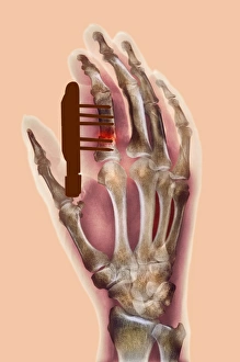
Background imageFractured Collection: Pinned finger fracture, X-ray

Pinned finger fracture, X-ray
Pinned broken finger. Coloured X-ray of a pinned fracture (red area) of the index finger. Five metal pins (dark brown) are holding the bone (the first phalanx)


All Professionally Made to Order for Quick Shipping

"Fractured: A Visual Journey of Broken Bones and Historical Moments" The X-ray reveals a broken wrist bone, a painful reminder of an unfortunate accident. Osteoporotic bone captured under SEM, showcasing the fragility that comes with age. An X-ray unveils a fractured ankle, highlighting the resilience needed for recovery. Lisfranc fracture caught on X-ray, reminding us of the intricate nature of foot injuries. Double fracture to the leg exposed by an X-ray, emphasizing the challenges faced in rehabilitation. The jawbone's fracture depicted through an X-ray image, illustrating both physical and emotional pain endured. A plaster cast encases a broken arm as seen in the X-ray, symbolizing healing and protection. Pinned broken leg showcased as medical intervention aids in mending shattered bones. "The Conquest of the Air, " painted in 1913 on canvas, reminds us that even great achievements can be marred by fractures within society itself. Bovie tool used to cut through retincaculum and clean up femur after Displaced patellar knee injury highlights modern medical advancements aiding recovery from fractures. Boy scouts exploring Cairo amidst ancient wonders signify youthful adventures despite potential risks like fractures along their journey Blitz aftermath captured on Standish Road in Hammersmith during WW2 serves as a stark reminder that war brings not only destruction but also countless fractures to lives and communities. In this captivating collection of images ranging from medical scans to historical moments, "Fractured" explores both literal and metaphorical breaks - from bones needing healing to societies torn apart by conflict or age-old divisions.