Fraud Blocker Skip to main content

Fractured Collection (page 3)

"Fractured: A Visual Journey of Broken Bones and Historical Moments" The X-ray reveals a broken wrist bone, a painful reminder of an unfortunate accident

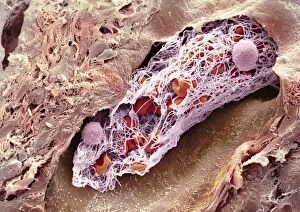
Background imageFractured Collection: Blood clot, SEM C014 / 0381

Blood clot, SEM C014 / 0381
Blood clot. Coloured scanning electron micrograph (SEM) of a blood clot (thrombus) in an arteriole (small blood vessel) of a salivary gland

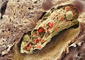
Background imageFractured Collection: Blood clot, SEM C014 / 0380

Blood clot, SEM C014 / 0380
Blood clot. Coloured scanning electron micrograph (SEM) of a blood clot (thrombus) in an arteriole (small blood vessel) of a salivary gland

Background imageFractured Collection: Fractured hip, artwork

Fractured hip, artwork
Fractured hip. Computer artwork of a fracture in the head of the femur (thigh bone)

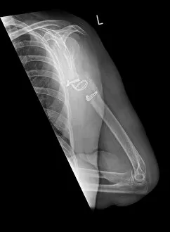
Background imageFractured Collection: Fractured and dislocated shoulder, X-ray C016 / 6607

Fractured and dislocated shoulder, X-ray C016 / 6607
Fractured and dislocated shoulder. Coloured X-ray of the shoulder of a 67 year old patient who has fallen and fractured the neck of the humerus (upper arm bone) and dislocated the shoulder

Background imageFractured Collection: Fractured and dislocated shoulder, X-ray C016 / 6606

Fractured and dislocated shoulder, X-ray C016 / 6606
Fractured and dislocated shoulder. Coloured X-ray of the shoulder of a 67 year old patient who has fallen and fractured the neck of the humerus (upper arm bone) and dislocated the shoulder

Background imageFractured Collection: Pinned leg fracture, X-ray C016 / 6586

Pinned leg fracture, X-ray C016 / 6586
Pinned leg fracture. X-ray of the leg of a 33 year old patient that has been externally fixed after a skiing accident. Both the tibia (shine bone, upper bone) and fibula (calf bone)

Background imageFractured Collection: Fractured pelvis, CT scan C016 / 6584

Fractured pelvis, CT scan C016 / 6584
Fractured pelvis. Coloured computed tomography (CT) scan of the pelvis of a 56 year old that was in a road traffic accident

Background imageFractured Collection: Pinned leg fracture, X-ray C016 / 6585

Pinned leg fracture, X-ray C016 / 6585
Pinned leg fracture. X-ray of the leg of a 33 year old patient that has been externally fixed after a skiing accident. Both the tibia (shine bone, upper bone) and fibula (calf bone)

Background imageFractured Collection: Q fever bacteria, SEM C017 / 8303

Q fever bacteria, SEM C017 / 8303
Q fever bacteria. Coloured scanning electron micrograph (SEM) of a fracture through a vacuole in a Vero cell showing replicating Coxiella burnetii bacteria (yellow)

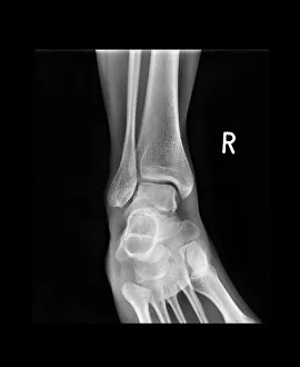
Background imageFractured Collection: Salter Harris fracture, X-ray C017 / 8025

Salter Harris fracture, X-ray C017 / 8025
Salter Harris fracture. X-ray of the ankle of a child patient with a displaced Salter Harris fracture of the ankle. Salter Harris fractures occur at the epiphyseal, or growth

Background imageFractured Collection: Ankle fracture with bone cyst, X-ray C017 / 8016

Ankle fracture with bone cyst, X-ray C017 / 8016
Ankle fracture. X-ray of the fractured ankle of a patient. The bone had been weakened by a cyst, a fluid-filled cavity

Background imageFractured Collection: Broken wrist, X-ray C017 / 8008

Broken wrist, X-ray C017 / 8008
Broken wrist. X-ray of the arm of an 84 year old female patient with a fractured wrist

Background imageFractured Collection: Pinned jaw fracture, X-ray C017 / 7982

Pinned jaw fracture, X-ray C017 / 7982
Pinned jaw fracture. X-ray of the head of a 22 year old male patient who has had a fractured mandible (lower jaw bone) pinned (lower left)

Background imageFractured Collection: Pinned broken ankle, X-ray C017 / 7978

Pinned broken ankle, X-ray C017 / 7978
Pinned broken ankle. X-ray of the ankle of a 32 year old male that has been pinned after a parachuting accident. The ankle was dislocated between the tibia (shin bone)

Background imageFractured Collection: Fractured and dislocated ankle, X-ray C017 / 7976

Fractured and dislocated ankle, X-ray C017 / 7976
Fractured and dislocated ankle. X-ray of the ankle of a 32 year old male who has been in a parachuting accident. The ankle is dislocated between the tibia (shin bone)

Background imageFractured Collection: Infected malunited fracture, X-ray C017 / 7972

Infected malunited fracture, X-ray C017 / 7972
Infected malunited fracture. X-ray of the arm of a 65 year old female patient who has a bone infection after the nonunion of a fracture of the humerus (upper arm bone)

Background imageFractured Collection: Thyroid gland, SEM C015 / 6419

Thyroid gland, SEM C015 / 6419
Thyroid gland. Coloured scanning electron micrograph (SEM) of a fracture through the thyroid gland revealing several follicles (green and blue). Between the follicles is connective tissue (pink)

Background imageFractured Collection: Ankle fracture with bone cyst, X-ray C017 / 7862

Ankle fracture with bone cyst, X-ray C017 / 7862
Ankle fracture. X-ray of the fractured ankle of a patient. The bone had been weakened by a cyst, a fluid-filled cavity

Background imageFractured Collection: Ankle bone fracture, X-ray C017 / 7809

Ankle bone fracture, X-ray C017 / 7809
Ankle bone fracture. X-ray of the ankle of a patient with a fractured talus (ankle bone)

Background imageFractured Collection: Ankle bone fracture, X-ray C017 / 7808

Ankle bone fracture, X-ray C017 / 7808
Ankle bone fracture. X-ray of the ankle of a patient with a fractured talus (ankle bone, lower centre)

Background imageFractured Collection: Broken elbow, X-ray C017 / 7750

Broken elbow, X-ray C017 / 7750
Broken elbow. X-ray of the arm of a 44 year old female patient with a broken elbow

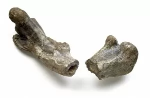
Background imageFractured Collection: Dryosaurus dinosaur, fossil thigh bone C016 / 5036

Dryosaurus dinosaur, fossil thigh bone C016 / 5036
Dryosaurus dinosaur, fossil thigh bone, showing the hollow bone structure. This fragmented femur is from Dryosaurus, a fast-running herbivorous dinosaur that lived during the Late Jurassic period

Background imageFractured Collection: Desiccation cracks in mud, Ecuador C013 / 8854

Desiccation cracks in mud, Ecuador C013 / 8854
Desiccation cracks in mud. View over cracked mud in a dry pond at the top of the beach. Photographed in Manabi Province, Ecuador

Background imageFractured Collection: Desiccation cracks in mud, Ecuador C013 / 8853

Desiccation cracks in mud, Ecuador C013 / 8853
Desiccation cracks in mud. Aerial view over cracked mud in a dry pond at the top of the beach. Photographed in Manabi Province, Ecuador

Background imageFractured Collection: Blood-filled artery, SEM C013 / 7117

Blood-filled artery, SEM C013 / 7117
Blood-filled artery. Coloured scanning electron micrograph (SEM) of a section through an artery (dark brown) in muscle tissue (light brown), showing the red blood cells (erythrocytes, centre) inside

Background imageFractured Collection: Blood-filled artery, SEM C013 / 7118

Blood-filled artery, SEM C013 / 7118
Blood-filled artery. Coloured scanning electron micrograph (SEM) of a section through an artery (centre) in muscle tissue, showing the red blood cells (erythrocytes, red) inside

Background imageFractured Collection: Blood-filled artery, SEM C013 / 7116

Blood-filled artery, SEM C013 / 7116
Blood-filled artery. Coloured scanning electron micrograph (SEM) of a section through an artery (dark pink, centre) in muscle tissue (purple), showing the red blood cells (erythrocytes, red) inside

Background imageFractured Collection: Hip fracture surgery C013 / 4960

Hip fracture surgery C013 / 4960
Hip fracture surgery. Surgeons operating on a patient during surgery to fix a hip fracture using a cannulated hip screw (CHS)

Background imageFractured Collection: Hip fracture surgery C013 / 4959

Hip fracture surgery C013 / 4959
Hip fracture surgery. Surgeons operating on a patient during surgery to fix a hip fracture using a cannulated hip screw (CHS)

Background imageFractured Collection: Hip fracture surgery C013 / 4958

Hip fracture surgery C013 / 4958
Hip fracture surgery. Surgical tools laid out ready for surgery to fix a hip fracture using a cannulated hip screw (CHS)

Background imageFractured Collection: Hip fracture surgery C013 / 4945

Hip fracture surgery C013 / 4945
Hip fracture surgery. Close up of a cannula in the hand of a patient undergoing surgery to fix a hip fracture using a cannulated hip screw (CHS)

Background imageFractured Collection: Hip fracture surgery C013 / 4940

Hip fracture surgery C013 / 4940
Hip fracture surgery. Surgeons operating on a patient during surgery to fix a hip fracture using a cannulated hip screw (CHS)

Background imageFractured Collection: Hip fracture surgery C013 / 4944

Hip fracture surgery C013 / 4944
Hip fracture surgery. Close up of a cannula in the leg of a patient undergoing surgery to fix a hip fracture using a cannulated hip screw (CHS)

Background imageFractured Collection: Hip fracture surgery C013 / 4943

Hip fracture surgery C013 / 4943
Hip fracture surgery. Close up of a surgeon closing an incision at the end of surgery to fix a hip fracture using a cannulated hip screw (CHS)

Background imageFractured Collection: Hip fracture surgery C013 / 4941

Hip fracture surgery C013 / 4941
Hip fracture surgery. Close up of surgeons operating on a patient during surgery to fix a hip fracture using a cannulated hip screw (CHS)

Background imageFractured Collection: Hip fracture surgery C013 / 4942

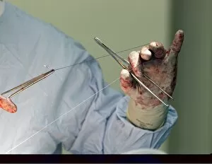
Hip fracture surgery C013 / 4942
Hip fracture surgery. Close up of a surgeon holding thread while stitching an incision at the end of surgery to fix a hip fracture using a cannulated hip screw (CHS)

Background imageFractured Collection: Hip fracture surgery C013 / 4939

Hip fracture surgery C013 / 4939
Hip fracture surgery. Close up of surgeons operating on a patient during surgery to fix a hip fracture using a cannulated hip screw (CHS)

Background imageFractured Collection: Hip fracture surgery C013 / 4938

Hip fracture surgery C013 / 4938
Hip fracture surgery. Close up of surgeons operating on a patient during surgery to fix a hip fracture using a cannulated hip screw (CHS)

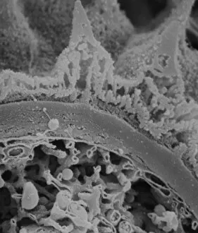
Background imageFractured Collection: Broken egg shell, SEM

Broken egg shell, SEM
Broken egg shell, coloured scanning electron micrograph (SEM). Hard-shelled eggs are produced by birds and some reptiles. The shell protects the embryo inside

Background imageFractured Collection: Egg shell membrane, SEM

Egg shell membrane, SEM
Egg shell membrane, coloured scanning electron micrograph (SEM). Broken egg shell, showing details of the membrane (across bottom) that is on the inside of the shell wall

Background imageFractured Collection: Blitz in London -- Standish Road, Hammersmith, WW2

Blitz in London -- Standish Road, Hammersmith, WW2
Blitz in London -- repairing a fractured water main in Standish Road, King Street, Hammersmith, 4 September 1940. A bomb-damaged house can be seen, top left

Background imageFractured Collection: Fractured pollen grain

Fractured pollen grain
Scanning electron microscope (SEM) image showing a fractured pollen grain

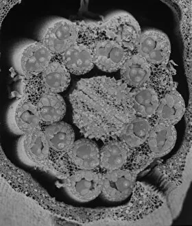
Background imageFractured Collection: Fractured anther

Fractured anther
Scanning electron microscope (SEM) image showing a fractured anther, otherwise known as the sac, which contains the pollen in the male sex organs (stamens)

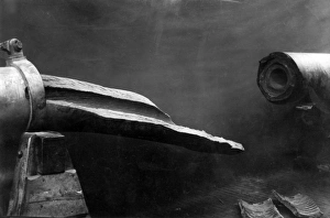
Background imageFractured Collection: Fractured barrel of German Paris Gun, WW1

Fractured barrel of German Paris Gun, WW1
The fractured barrel of the German Paris Gun (also known as the Emperor William Gun), a long-range seige gun designed and manufactured by Krupp, used towards the end of the First World War

Background imageFractured Collection: Dog with Jaw Splint

Dog with Jaw Splint
A fixation splint for a fractured jaw, the latest development. Date: circa 1960

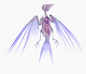
Background imageFractured Collection: Swallow, X-ray

Swallow, X-ray
Swallow. Coloured X-ray of a swallow (family Hirundidae). The swallow spends much of its time on the wing, and it is well adapted for energy- efficient flight; its hollow bones

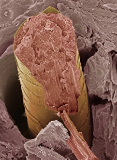
Background imageFractured Collection: Hair, SEM

Hair, SEM
Hair fracture. Coloured scanning electron micrograph (SEM) of a fractured hair (yellow and pink). The surrounding layer of facial skin (brown) has a typically scale-like surface

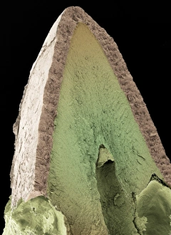
Background imageFractured Collection: Cats tooth, SEM

Cats tooth, SEM
Cats tooth. Coloured scanning electron micrograph (SEM) of a vertical fracture through a canine tooth of a domestic cat (Felis catus)


All Professionally Made to Order for Quick Shipping

"Fractured: A Visual Journey of Broken Bones and Historical Moments" The X-ray reveals a broken wrist bone, a painful reminder of an unfortunate accident. Osteoporotic bone captured under SEM, showcasing the fragility that comes with age. An X-ray unveils a fractured ankle, highlighting the resilience needed for recovery. Lisfranc fracture caught on X-ray, reminding us of the intricate nature of foot injuries. Double fracture to the leg exposed by an X-ray, emphasizing the challenges faced in rehabilitation. The jawbone's fracture depicted through an X-ray image, illustrating both physical and emotional pain endured. A plaster cast encases a broken arm as seen in the X-ray, symbolizing healing and protection. Pinned broken leg showcased as medical intervention aids in mending shattered bones. "The Conquest of the Air, " painted in 1913 on canvas, reminds us that even great achievements can be marred by fractures within society itself. Bovie tool used to cut through retincaculum and clean up femur after Displaced patellar knee injury highlights modern medical advancements aiding recovery from fractures. Boy scouts exploring Cairo amidst ancient wonders signify youthful adventures despite potential risks like fractures along their journey Blitz aftermath captured on Standish Road in Hammersmith during WW2 serves as a stark reminder that war brings not only destruction but also countless fractures to lives and communities. In this captivating collection of images ranging from medical scans to historical moments, "Fractured" explores both literal and metaphorical breaks - from bones needing healing to societies torn apart by conflict or age-old divisions.