
Fractured Collection (page 2)
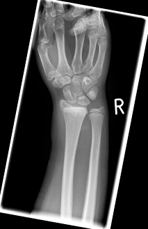
"Fractured: A Visual Journey of Broken Bones and Historical Moments" The X-ray reveals a broken wrist bone, a painful reminder of an unfortunate accident


All Professionally Made to Order for Quick Shipping
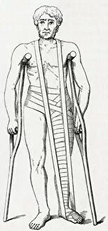
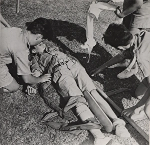
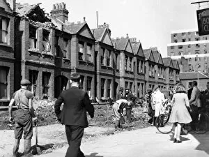
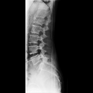
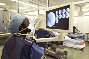
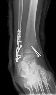
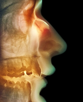
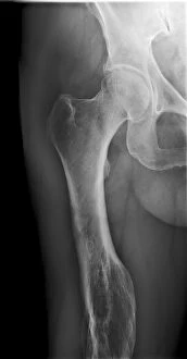
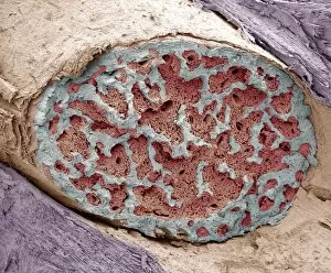
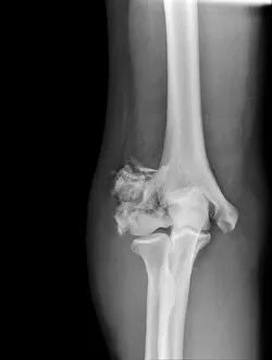
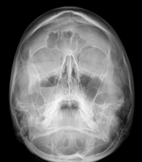
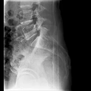
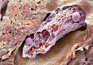
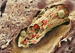
"Fractured: A Visual Journey of Broken Bones and Historical Moments" The X-ray reveals a broken wrist bone, a painful reminder of an unfortunate accident. Osteoporotic bone captured under SEM, showcasing the fragility that comes with age. An X-ray unveils a fractured ankle, highlighting the resilience needed for recovery. Lisfranc fracture caught on X-ray, reminding us of the intricate nature of foot injuries. Double fracture to the leg exposed by an X-ray, emphasizing the challenges faced in rehabilitation. The jawbone's fracture depicted through an X-ray image, illustrating both physical and emotional pain endured. A plaster cast encases a broken arm as seen in the X-ray, symbolizing healing and protection. Pinned broken leg showcased as medical intervention aids in mending shattered bones. "The Conquest of the Air, " painted in 1913 on canvas, reminds us that even great achievements can be marred by fractures within society itself. Bovie tool used to cut through retincaculum and clean up femur after Displaced patellar knee injury highlights modern medical advancements aiding recovery from fractures. Boy scouts exploring Cairo amidst ancient wonders signify youthful adventures despite potential risks like fractures along their journey Blitz aftermath captured on Standish Road in Hammersmith during WW2 serves as a stark reminder that war brings not only destruction but also countless fractures to lives and communities. In this captivating collection of images ranging from medical scans to historical moments, "Fractured" explores both literal and metaphorical breaks - from bones needing healing to societies torn apart by conflict or age-old divisions.