Fraud Blocker Skip to main content

Alpha Helix Collection (page 4)

The alpha helix, a fundamental structure in biology, plays a crucial role in various molecular processes

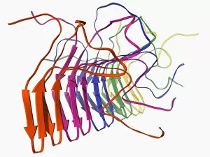
Background imageAlpha Helix Collection: Fungal prion protein F006 / 9644

Fungal prion protein F006 / 9644
Fungal prion protein. Molecular model of the amyloid form of the HET prion protein

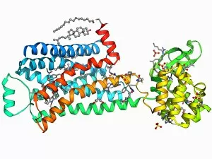
Background imageAlpha Helix Collection: Beta-2 adrenergic receptor molecule F006 / 9643

Beta-2 adrenergic receptor molecule F006 / 9643
Beta-2 adrenergic receptor. Molecular model of a human beta-2 adrenergic receptor bound to an antibody. Beta receptors respond to adrenalin, causing a sympathetic (fight or flight) response

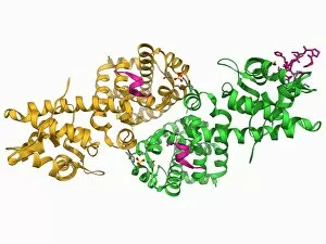
Background imageAlpha Helix Collection: Adenovirus protein and tumor suppressor F006 / 9641

Adenovirus protein and tumor suppressor F006 / 9641
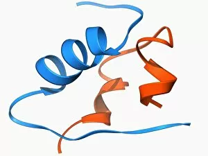
Adenovirus protein and tumour suppressor. Molecular model of the E1A protein from human adenovirus bound to a retinoblastoma tumour suppressor

Background imageAlpha Helix Collection: Beta-2 adrenergic receptor molecule F006 / 9640

Beta-2 adrenergic receptor molecule F006 / 9640
Beta-2 adrenergic receptor. Molecular model of a human beta-2 adrenergic receptor bound to an antibody. Beta receptors respond to adrenalin, causing a sympathetic (fight or flight) response

Background imageAlpha Helix Collection: E coli virulence factor F006 / 9639

E coli virulence factor F006 / 9639
E. coli virulence factor. Molecular model of the beta-domain of the EspP autotransporter protein from the bacterium Escherichia coli

Background imageAlpha Helix Collection: 70S ribosome, molecular model F006 / 9638

70S ribosome, molecular model F006 / 9638
70S ribosome. Molecular model of a 70S ribosome complex containing a Shine-Dalgarno helix, the point of mRNA (messenger ribonucleic acid) binding

Background imageAlpha Helix Collection: Metal-sensing RNA molecule F006 / 9636

Metal-sensing RNA molecule F006 / 9636
Metal-sensing RNA molecule. Molecular model of an M-box riboswitch, a length of RNA (ribonucleic acid) that regulates levels of metal ions in a cell

Background imageAlpha Helix Collection: Trypsin molecule F006 / 9634

Trypsin molecule F006 / 9634
Trypsin molecule. Molecular model of the digestive protease enzyme trypsin. Trypsin is released by the pancreas to break down proteins into smaller chains of amino acids

Background imageAlpha Helix Collection: PolyA polymerase and RNA F006 / 9635

PolyA polymerase and RNA F006 / 9635
Poly(A) polymerase and RNA. Molecular model of poly(A) polymerase complexed with RNA (ribonucleic acid) and ATP (adenosine triphosphate)

Background imageAlpha Helix Collection: Internal ribosome entry site F006 / 9631

Internal ribosome entry site F006 / 9631
Internal ribosome entry site. Molecular model of an internal ribosome entry site nucleotide sequence from the hepatitis C virus. This sequence is essential for the initiation of viral translation

Background imageAlpha Helix Collection: Trypsin molecule with inhibitor F006 / 9633

Trypsin molecule with inhibitor F006 / 9633
Trypsin molecule. Molecular model of the digestive protease enzyme beta-trypsin complexed with an inhibitor. Trypsin is released by the pancreas to break down proteins into smaller chains of amino

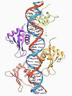
Background imageAlpha Helix Collection: Interferon regulatory factor molecule F006 / 9630

Interferon regulatory factor molecule F006 / 9630
Interferon regulatory factor. Molecular model of interferon regulatory factor 3 (IRF3, coils at right and left) bound to a DNA (deoxyribonucleic acid) molecule (red and blue)

Background imageAlpha Helix Collection: Wilms tumor suppressor bound to DNA F006 / 9632

Wilms tumor suppressor bound to DNA F006 / 9632
Wilms tumour suppressor bound to DNA. Molecular model of the zinc finger domain of the Wilms tumour suppressor protein bound to a strand of DNA (deoxyribonucleic acid)

Background imageAlpha Helix Collection: Lactose binding protein molecule F006 / 9629

Lactose binding protein molecule F006 / 9629
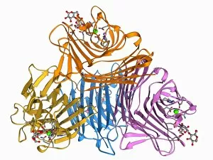
Lactose binding protein. Molecular model of a lectin protein from the peanut plant (Arachis hypogaea) bound to a lactose molecule

Background imageAlpha Helix Collection: MscS ion channel protein structure F006 / 9626

MscS ion channel protein structure F006 / 9626
MscS ion channel protein structure. Molecular model of a mechanosensitive channel of small conductance (MscS) from an Escherichia coli bacterium

Background imageAlpha Helix Collection: Transport inhibitor response 1 protein F006 / 9628

Transport inhibitor response 1 protein F006 / 9628
Transport inhibitor response 1 protein. Molecular model of the transport inhibitor response 1 protein bound to the plant hormone auxin. This protein is involved in auxin gene regulation

Background imageAlpha Helix Collection: Multidrug transporter molecule F006 / 9627

Multidrug transporter molecule F006 / 9627
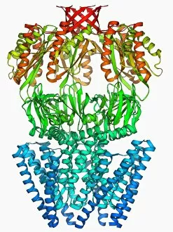
Multidrug transporter. Molecular model of the multidrug transporter Sav1866 from the bacterium Escherichia coli. This protein pumps drugs, including antibiotics, out of the bacterial cell

Background imageAlpha Helix Collection: Mengovirus capsid, molecular model F006 / 9617

Mengovirus capsid, molecular model F006 / 9617
Mengovirus capsid, molecular model. A capsid consists of subunits called capsomeres that self-assemble to form the shell seen here

Background imageAlpha Helix Collection: Insulin molecule F006 / 9625

Insulin molecule F006 / 9625
Insulin, molecular model. Insulin plays an important role in blood sugar regulation. It is released from the pancreas when blood sugar levels are high, for example after a meal

Background imageAlpha Helix Collection: MscL ion channel protein structure F006 / 9624

MscL ion channel protein structure F006 / 9624
MscL ion channel protein structure. Molecular model of a mechanosensitive channel of large conductance (MscL) from a Mycobacterium tuberculosis bacterium

Background imageAlpha Helix Collection: Endoplasmic reticulum chaperone protein F006 / 9623

Endoplasmic reticulum chaperone protein F006 / 9623
Endoplasmic reticulum chaperone protein. Molecular model of the endoplasmic reticulum chaperone protein GRP94. This protein is essential for the maturation of cell-surface display proteins

Background imageAlpha Helix Collection: RNA exosome complex, molecular model F006 / 9620

RNA exosome complex, molecular model F006 / 9620
RNA exosome complex, molecular model. This multi-protein complex functions to break up strands of RNA (ribonucleic acid, pink) during biochemical processes

Background imageAlpha Helix Collection: HIV antibody therapy, molecular model F006 / 9622

HIV antibody therapy, molecular model F006 / 9622
HIV antibody therapy. Molecular model of the interaction of the HIV surface protein gp120 (green) as it interacts with a human white blood cell surface protein (CD4)

Background imageAlpha Helix Collection: Rhomboid protease molecule F006 / 9621

Rhomboid protease molecule F006 / 9621
Rhomboid protease. Molecular model of the rhomboid protease enzyme GlpG from the bacterium Escherichia coli. Proteases are enzymes that break down proteins

Background imageAlpha Helix Collection: Kinesin motor protein F006 / 9619

Kinesin motor protein F006 / 9619
Kinesin motor protein. Molecular model of the ncd kinesin motor protein. Kinesin motor proteins transport vesicles containing intracellular cargo around the cell along microtubules

Background imageAlpha Helix Collection: Molecular motor protein F006 / 9618

Molecular motor protein F006 / 9618
Myosin molecular motor protein, molecular model. Motor proteins convert chemical energy into mechanical movements in response to specific chemical stimuli

Background imageAlpha Helix Collection: RNA editing enzyme F006 / 9615

RNA editing enzyme F006 / 9615
RNA editing enzyme, molecular model. This enzyme binds to double-stranded RNA (ribonucleic acid)

Background imageAlpha Helix Collection: Lysozyme molecule F006 / 9616

Lysozyme molecule F006 / 9616
Lysozyme, molecular model. Lysozymes are enzymes found in a wide range of biological fluids such as tears, saliva and milk. This lysozyme is from chicken egg white

Background imageAlpha Helix Collection: RNA-dependent RNA polymerase molecule F006 / 9611

RNA-dependent RNA polymerase molecule F006 / 9611
RNA-dependent RNA polymerase, molecular model. This enzyme catalyses the replication of RNA (ribonucleic acid) from an RNA template

Background imageAlpha Helix Collection: Titin muscle protein molecule F006 / 9612

Titin muscle protein molecule F006 / 9612
Titin muscle protein. Molecular model of two immunoglobulin-like domains from the giant muscle protein titin

Background imageAlpha Helix Collection: Integrin transmembrane domain F006 / 9614

Integrin transmembrane domain F006 / 9614
Integrin transmembrane domain, molecular model. Integrins are transmembrane cell adhesion receptors

Background imageAlpha Helix Collection: Cytochrome P450 and erythromycin F006 / 9610

Cytochrome P450 and erythromycin F006 / 9610
Cytochrome P450 and erythromycin. Molecular model of human cytochrome P450 complexed with the antibiotic erythromycin. This protein plays a crucial role in metabolism in animals (including humans)

Background imageAlpha Helix Collection: Growth factor receptor molecule F006 / 9613

Growth factor receptor molecule F006 / 9613
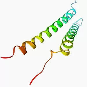
Growth factor receptor. Molecular model of the transmembrane segment of the ErbB2 growth factor receptor

Background imageAlpha Helix Collection: Nerve growth factor bound to receptor F006 / 9608

Nerve growth factor bound to receptor F006 / 9608
Nerve growth factor. Molecular model of human nerve growth factor bound to the TrkA receptor. NGF is a neurotrophin that acts on the development and function of nerves

Background imageAlpha Helix Collection: Viral RNA packaging signal complex F006 / 9609

Viral RNA packaging signal complex F006 / 9609
Viral RNA packaging signal complex. Molecular model of the muPsi RNA packaging signal complex from the Rous sarcoma vuris

Background imageAlpha Helix Collection: HIV reverse transcription enzyme F006 / 9606

HIV reverse transcription enzyme F006 / 9606
HIV reverse transcription enzyme. Molecular model of the reverse transcriptase enzyme (pink) found in HIV (the human immunodeficiency virus)

Background imageAlpha Helix Collection: Haemoglobin, molecular model F006 / 9604

Haemoglobin, molecular model F006 / 9604
Haemoglobin, molecular model. This is deoxyhaemoglobin, the molecule in its non-oxygen bound state. Haemoglobin transports oxygen around the body in red blood cells

Background imageAlpha Helix Collection: Rhomboid protease molecule F006 / 9607

Rhomboid protease molecule F006 / 9607
Rhomboid protease. Molecular model of the rhomboid protease enzyme GlpG from the bacterium Escherichia coli. Proteases are enzymes that break down proteins

Background imageAlpha Helix Collection: Glutamine synthetase enzyme F006 / 9598

Glutamine synthetase enzyme F006 / 9598
Glutamine synthetase enzyme, molecular model. This ligase enzyme forms chemical bonds between molecules. It plays an important role in the metabolism of nitrogen by catalysing the condensation of

Background imageAlpha Helix Collection: Insulin molecule F006 / 9605

Insulin molecule F006 / 9605
Insulin molecule. Molecular model of the hormone insulin. Insulin consists of two peptide chains, A and B, which are linked by disulphide bridges

Background imageAlpha Helix Collection: 3-hydroxyacyl-CoA dehydrogenase F006 / 9602

3-hydroxyacyl-CoA dehydrogenase F006 / 9602
3-hydroxyacyl-CoA dehydrogenase, molecular model. This enzyme is found in human heart tissue, and catalyses a reaction that is part of the beta-oxidation pathway

Background imageAlpha Helix Collection: Hepatitis B virus capsid, molecular model F006 / 9594

Hepatitis B virus capsid, molecular model F006 / 9594
Hepatitis B virus capsid, molecular model. This virus, transmitted through infected bodily fluids or blood, causes the disease hepatitis B in humans, leading to acute liver inflammation

Background imageAlpha Helix Collection: Thrombin protein, molecular model F006 / 9603

Thrombin protein, molecular model F006 / 9603
Thrombin protein, molecular model. Thrombin is an enzyme involved in the blood coagulation (clotting) process. It converts fibrinogen (a soluble plasma glycoprotein synthesised in the liver)

Background imageAlpha Helix Collection: Haemoglobin S, molecular model F006 / 9601

Haemoglobin S, molecular model F006 / 9601
Haemoglobin S. Molecular model of the mutant form of haemoglobin (haemoglobin S) that causes sickle cell anaemia. This is deoxyhaemoglobin S, the molecule in its non-oxygen bound state

Background imageAlpha Helix Collection: RNA-editing enzyme, molecular model F006 / 9599

RNA-editing enzyme, molecular model F006 / 9599
RNA-editing enzyme. Molecular model of a left-handed, RNA double helix (Z-RNA, centre) bound by the Z alpha domain of the human RNA-editing enzyme ADAR1 (double-stranded RNA adenosine deaminase)

Background imageAlpha Helix Collection: Protein kinase regulatory subunit F006 / 9600

Protein kinase regulatory subunit F006 / 9600
Protein kinase regulatory subunit. Molecular model of a regulatory subunit from cAMP-dependent protein kinase bound to. This enzyme is also known as protein kinase A (PKA)

Background imageAlpha Helix Collection: Cyanobacterial circadian clock protein F006 / 9595

Cyanobacterial circadian clock protein F006 / 9595
Cyanobacterial circadian clock protein, molecular model. This protein is a kinase known as KaiC. Its structure is a hexamer

Background imageAlpha Helix Collection: Programmed cell death protein molecule F006 / 9597

Programmed cell death protein molecule F006 / 9597
Human programmed cell death protein 4, molecular model. This protein is involved in apoptosis (programmed cell death)


All Professionally Made to Order for Quick Shipping

The alpha helix, a fundamental structure in biology, plays a crucial role in various molecular processes. From DNA transcription to protein synthesis, this intricate arrangement is found throughout the biological world. In the realm of genetics, the alpha helix participates in DNA transcription by aiding in the unwinding and separation of strands. Its elegant spiral shape allows for efficient reading and copying of genetic information. When it comes to proteins, the alpha helix serves as a secondary structure that contributes to their stability and function. Visualized through stunning artwork or molecular models, its coiled form adds strength and flexibility to these vital biomolecules. One example where we can observe this remarkable structure is within the nucleosome molecule. Here, DNA wraps around histone proteins forming tight coils resembling beads on a string – with each bead representing an alpha helix. Another instance occurs within bacterial ribosomes responsible for protein synthesis. The presence of multiple alpha helices enables precise positioning of molecules during translation – ensuring accurate assembly of amino acids into functional proteins. Viruses also exploit this structural motif; one such case being HIV reverse transcription enzyme. This enzyme utilizes an alpha helical region to convert viral RNA into DNA – a critical step in viral replication. Similarly, hepatitis C virus enzyme employs an intricate network of alpha helices depicted by molecular models. These structures aid in catalyzing chemical reactions necessary for viral survival and proliferation. Moving beyond viruses, manganese superoxide dismutase enzyme showcases how nature harnesses the power of the alpha helix for antioxidant defense mechanisms within cells. Its tightly wound coils protect against harmful free radicals that can damage cellular components. Alpha-helical motifs are not limited to enzymes alone but extend to larger molecules like human serum albumin or Argonaute protein involved in gene regulation pathways. Their well-defined arrangements contribute significantly to their respective functions within our bodies' complex systems.