Fraud Blocker Skip to main content

Alpha Helix Collection (page 15)

The alpha helix, a fundamental structure in biology, plays a crucial role in various molecular processes

Background imageAlpha Helix Collection: GST enzyme conferring DDT resistance

GST enzyme conferring DDT resistance
Glutathione S-transferase (GST) enzyme from a malaria-carrying strain of mosquito, Anopheles gambiae, molecular model. GSTs detoxify foreign substances in the body of the mosquito

Background imageAlpha Helix Collection: Testosterone bound to receptor, artwork

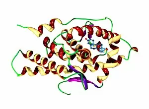
Testosterone bound to receptor, artwork
Testosterone bound to receptor. Molecular model of a molecule of the male sex hormone testosterone (ball-and-stick primary structure) bound to a human androgen receptor (secondary structure)

Background imageAlpha Helix Collection: B domain of staphylococcal protein A

B domain of staphylococcal protein A
B domain of Staphylococcus aureus protein A, molecular model. Protein A is a surface factor found on the membrane of S. aureus bacteria

Background imageAlpha Helix Collection: Cobra venom action, molecular model

Cobra venom action, molecular model
Cobra (Naja sp.) venom action. Molecular model showing top (upper centre) and side (lower centre) views of the secondary structure of an alpha-cobratoxin (snake venom protein)

Background imageAlpha Helix Collection: RNA polymerase from Norwalk virus

RNA polymerase from Norwalk virus
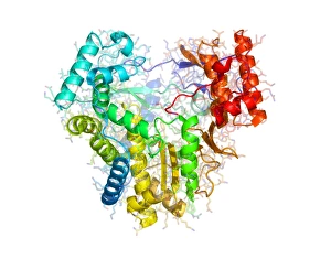
RNA polymerase enzyme from Norwalk virus, molecular model. This enzyme makes RNA (ribonucleic acid) from the RNA template within the virus

Background imageAlpha Helix Collection: RNA polymerase from rabies virus

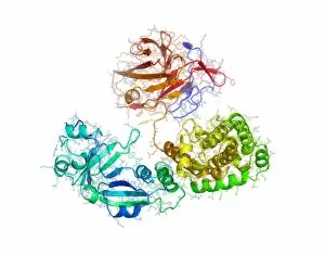
RNA polymerase from rabies virus, molecular model. This is a single subunit from an enzyme, involved in the replication of the rabies viruss RNA after it has infected a host cell

Background imageAlpha Helix Collection: Phosphofructokinase bacterial enzyme

Phosphofructokinase bacterial enzyme
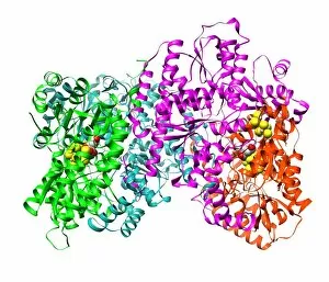
Phosphofructokinase enzyme, molecular model. This enzyme is from the spirochaete bacteria Borrelia burgdorferi, which causes Lyme disease

Background imageAlpha Helix Collection: Diphtheria toxin structure

Diphtheria toxin structure
Diphtheria toxin, molecular model. This model shows the toxin produced by the bacterium Corynebacterium diphtheriae, the cause of diphtheria

Background imageAlpha Helix Collection: Hepatitis C virus RNA polymerase enzyme

Hepatitis C virus RNA polymerase enzyme
Hepatitis C virus enzyme, molecular model. This protein, called NS5B, forms the active site of the viruss RNA-dependent RNA polymerase enzyme

Background imageAlpha Helix Collection: Parathyroid hormone, molecular model

Parathyroid hormone, molecular model
Parathyroid hormone. Molecular model showing the primary structure (spheres) and secondary structure (coils) of parathyroid hormone (PTH), or parathormone

Background imageAlpha Helix Collection: Protein from measles virus

Protein from measles virus
Proteins from the measles virus, molecular model. A domain of the viruss P protein (upper, blue, green and yellow) is seen here in complex with part of the N protein (lower, red)

Background imageAlpha Helix Collection: Phenylbutazone anti-inflammatory molecule

Phenylbutazone anti-inflammatory molecule
Phenylbutazone anti-inflammatory drug. Molecular model showing the secondary structure of the non-steroidal anti-inflammatory drug (NSAID) phenylbutazone

Background imageAlpha Helix Collection: THG anabolic steroid and receptor

THG anabolic steroid and receptor
THG bound to receptor. Molecular model of a molecule of the anabolic steroid drug tetrahydrogestrinone (THG, ball-and-stick primary structure) bound to a human androgen receptor (secondary structure)

Background imageAlpha Helix Collection: HIV antibody therapy

HIV antibody therapy. Molecular model of the interaction of the HIV surface protein gp120 (green, lower right) as it interacts with a human white blood cell surface protein (CD4, blue)

Background imageAlpha Helix Collection: Cholesterol enzyme affected by a drug

Cholesterol enzyme affected by a drug
Cholesterol enzyme being affected by a drug. Molecular model of the shape of the human enzyme Hmg-Coa reductase interacting (complexed) with the anti-cholesterol drug Fluvastatin (not seen)

Background imageAlpha Helix Collection: HIV enzyme being affected by a drug

HIV enzyme being affected by a drug. Molecular model of HIVs reverse transcriptase enzyme as it interacts with a drug (not seen)

Background imageAlpha Helix Collection: Erythropoietin molecule bound to receptor

Erythropoietin molecule bound to receptors. Computer model of a molecule of erythropoietin (EPO) (orange) bound to two extracellular EPO receptors (pink and purple)

Background imageAlpha Helix Collection: Insulin hormone, molecular model

Insulin hormone, molecular model
Insulin hormone. Molecular model of the bovine form of the hormone insulin, produced by the pancreas in mammals to aid the body in metabolising sugars

Background imageAlpha Helix Collection: Thrombin protein, secondary structure

Thrombin protein, secondary structure
Thrombin protein, computer model. Thrombin is a protein involved in the blood coagulation (clotting) process. It converts fibrinogen (a soluble plasma glycoprotein synthesised in the liver)

Background imageAlpha Helix Collection: GAGA transcription factor molecule

GAGA transcription factor molecule. Molecular model showing the primary (rods) and secondary (helices) structure of GAGA factor (green and blue)

Background imageAlpha Helix Collection: Cytochrome P450 protein, molecular model

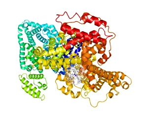
Cytochrome P450 protein, molecular model. This protein plays a crucial role in metabolism in animals (including humans), fungi, plants and bacteria

Background imageAlpha Helix Collection: Src protein domain

Src protein domain. Computer model showing the primary (rods) and secondary (alpha helix, blue and beta sheets, purple) structures of the Src homology domain 3 (SH3)

Background imageAlpha Helix Collection: Integration host factor and DNA

Integration host factor and DNA. Computer model of integration host factor (IHF, centre) bound to a molecule of DNA (deoxyribonucleic acid, semi- circle). The secondary structure of IHF is shown

Background imageAlpha Helix Collection: Human rhinovirus capsid proteins

Human rhinovirus capsid proteins, molecular model. These are proteins from the capsid (outer protein coat) of rhinovirus 14. Rhinoviruses are responsible for causing about 50% of common colds

Background imageAlpha Helix Collection: Haemagglutinin from bird flu virus

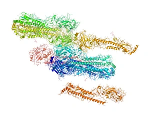
Haemagglutinin from bird flu virus, molecular model. This protein, H5, is found on the surface of the bird flu virus H5N1

Background imageAlpha Helix Collection: Enzyme from a sulphur-reducing bacterium

Enzyme from a sulphur-reducing bacterium. Molecular model of the enzyme aldehyde oxidoreductase from the Desulfovibrio gigas sulphur-reducing bacterium

Background imageAlpha Helix Collection: Yeast enzyme, molecular model

Yeast enzyme, molecular model
Yeast enzyme. Molecular model of an enzyme from Saccharomyces cerevisiae (Bakers yeast). This enzyme is 20S proteasome. A proteasome is a complex type of proteinase (protein-digesting enzyme)

Background imageAlpha Helix Collection: Fibroblast growth factor receptor 2

Fibroblast growth factor receptor 2 (FGFR2). Molecular models of the secondary structure (top) and the tertiary structure (bottom) of FGFR2

Background imageAlpha Helix Collection: Single stranded DNA-binding protein

Single stranded DNA-binding protein (SSBP). Molecular model showing the secondary and tertiary structures of a protein that binds to the single stranded DNA (deoxyribonucleic acid)

Background imageAlpha Helix Collection: Nitrogenase protein, molecular model

Nitrogenase protein, molecular model
Nitrogenase protein. Molecular model of the MoFe protein, one of two proteins (MoFe and Fe) that combine to form the enzyme nitrogenase


All Professionally Made to Order for Quick Shipping

The alpha helix, a fundamental structure in biology, plays a crucial role in various molecular processes. From DNA transcription to protein synthesis, this intricate arrangement is found throughout the biological world. In the realm of genetics, the alpha helix participates in DNA transcription by aiding in the unwinding and separation of strands. Its elegant spiral shape allows for efficient reading and copying of genetic information. When it comes to proteins, the alpha helix serves as a secondary structure that contributes to their stability and function. Visualized through stunning artwork or molecular models, its coiled form adds strength and flexibility to these vital biomolecules. One example where we can observe this remarkable structure is within the nucleosome molecule. Here, DNA wraps around histone proteins forming tight coils resembling beads on a string – with each bead representing an alpha helix. Another instance occurs within bacterial ribosomes responsible for protein synthesis. The presence of multiple alpha helices enables precise positioning of molecules during translation – ensuring accurate assembly of amino acids into functional proteins. Viruses also exploit this structural motif; one such case being HIV reverse transcription enzyme. This enzyme utilizes an alpha helical region to convert viral RNA into DNA – a critical step in viral replication. Similarly, hepatitis C virus enzyme employs an intricate network of alpha helices depicted by molecular models. These structures aid in catalyzing chemical reactions necessary for viral survival and proliferation. Moving beyond viruses, manganese superoxide dismutase enzyme showcases how nature harnesses the power of the alpha helix for antioxidant defense mechanisms within cells. Its tightly wound coils protect against harmful free radicals that can damage cellular components. Alpha-helical motifs are not limited to enzymes alone but extend to larger molecules like human serum albumin or Argonaute protein involved in gene regulation pathways. Their well-defined arrangements contribute significantly to their respective functions within our bodies' complex systems.