Canvas Print > Popular Themes > Human Body
Canvas Print : Heart structure


![]()
Canvas Prints from Science Photo Library
Heart structure
Artwork of the internal structure of the heart. On the left, the right atrium (top cavity) and right ventriculum (bottom) receive deoxygenated blood from the body through the superior and inferior vena cava, coloured blue. From the right ventriculum the blood flows through the pulmonary arteries (not visible) to the lungs. Oxygenated blood returns to the left atrium and ventriculum and is distributed to the body through the red ascending aorta. The yellow feature at the base of the superior vena cava, embedded in blue nerve endings, is the sinuatrial node where the impulse for the heart contraction originates
Science Photo Library features Science and Medical images including photos and illustrations
Media ID 6420778
© FRANCIS LEROY, BIOCOSMOS/SCIENCE PHOTO LIBRARY
Atria Atrium Blood System Body Cardiology Circulatory Cross Section Ventricle Ventricles Circulation
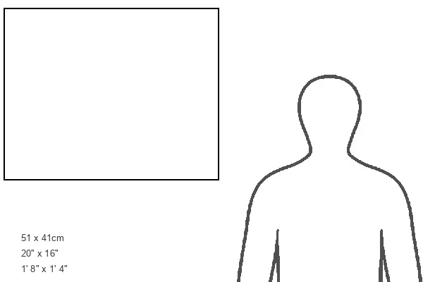
20"x16" (51x41cm) Canvas Print
Bring the wonders of science into your home with Media Storehouse's Canvas Prints. This stunning artwork, featuring the intricate internal structure of the heart from Science Photo Library, is a testament to the beauty and complexity of the human body. The right atrium and ventricle, clearly depicted on the left, receive deoxygenated blood from the body, ready to be pumped through the circulatory system. Order now and let this captivating print serve as a daily reminder of the marvels of science and the intricacies of the heart.
Delivered stretched and ready to hang our premium quality canvas prints are made from a polyester/cotton blend canvas and stretched over a 1.25" (32mm) kiln dried knot free wood stretcher bar. Packaged in a plastic bag and secured to a cardboard insert for safe transit.
Canvas Prints add colour, depth and texture to any space. Professionally Stretched Canvas over a hidden Wooden Box Frame and Ready to Hang
Estimated Product Size is 50.8cm x 40.6cm (20" x 16")
These are individually made so all sizes are approximate
Artwork printed orientated as per the preview above, with landscape (horizontal) orientation to match the source image.
EDITORS COMMENTS
This print showcases the intricate internal structure of the heart, beautifully depicted as a work of art. On the left side, we observe the right atrium at the top and right ventricle at the bottom, both responsible for receiving deoxygenated blood from our body through the superior and inferior vena cava, represented by shades of blue. The unseen pulmonary arteries transport this blood to our lungs for oxygenation. The image also highlights how oxygenated blood returns to the heart's left atrium and ventricle before being distributed throughout our body via the vibrant red ascending aorta. At its base lies a fascinating yellow feature known as the sinuatrial node - an essential part where impulses for heart contractions originate. This tiny node is surrounded by delicate blue nerve endings, adding another layer of complexity to this remarkable organ. With its emphasis on healthy anatomy and normal circulation within our bodies, this illustration provides us with valuable insights into human cardiology and circulatory systems. It reminds us of just how vital these structures are in maintaining overall well-being. Captured by Science Photo Library, renowned for their exceptional visual representations in various scientific fields, this print serves as a testament to their expertise in capturing not only scientific accuracy but also artistic beauty within medical imagery.
MADE IN THE USA
Safe Shipping with 30 Day Money Back Guarantee
FREE PERSONALISATION*
We are proud to offer a range of customisation features including Personalised Captions, Color Filters and Picture Zoom Tools
SECURE PAYMENTS
We happily accept a wide range of payment options so you can pay for the things you need in the way that is most convenient for you
* Options may vary by product and licensing agreement. Zoomed Pictures can be adjusted in the Cart.