Fraud Blocker Skip to main content

Home > Science Photo Library > Images Dated > 2011 > April > 27 Apr 2011

Images Dated 27th April 2011

Choose a picture from our Images Dated 27th April 2011 Collection for your Wall Art and Photo Gifts

32 items

We are proud to offer this selection in partnership with Science Photo Library

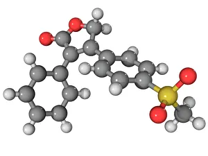
Background imageImages Dated 27th April 2011: Vioxx drug molecule

Vioxx drug molecule
Vioxx. Molecular model of the non-steroidal anti-inflammatory drug rofecoxib, which was marketed as Vioxx. This drug was withdrawn from use in 2004 due to concerns over increased heart attack

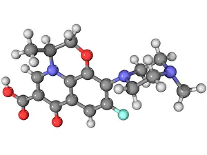
Background imageImages Dated 27th April 2011: Levofloxacin antibiotic molecule

Levofloxacin antibiotic molecule
Levofloxacin antibiotic, molecular model. Atoms are represented as spheres and are colour-coded: carbon (grey), hydrogen (white), nitrogen (dark blue), oxygen (red) and fluorine (light blue)

Background imageImages Dated 27th April 2011: Human chromosome 10, SEM

Human chromosome 10, SEM
Human chromosome 10, coloured scanning electron micrograph (SEM). Chromosomes are a packaged form of the genetic material DNA (deoxyribonucleic acid)

Background imageImages Dated 27th April 2011: Human chromosome 16, SEM

Human chromosome 16, SEM
Human chromosome 16, coloured scanning electron micrograph (SEM). Chromosomes are a packaged form of the genetic material DNA (deoxyribonucleic acid)

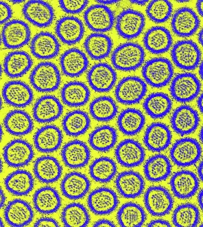
Background imageImages Dated 27th April 2011: Intestinal microvilli, TEM

Intestinal microvilli, TEM
Intestinal microvilli, coloured transmission electron micrograph (TEM). The microvilli are finger-like projections (seen here in circular cross-section)

Background imageImages Dated 27th April 2011: Plutonium crystal structure

Plutonium crystal structure, molecular model

Background imageImages Dated 27th April 2011: Vardenafil erectile dysfunction drug

Vardenafil erectile dysfunction drug
Vardenafil, molecular model. This erectile dysfunction drug is marketed as Levitra. Atoms are represented as spheres and are colour-coded: carbon (grey), hydrogen (white), oxygen (red)

Background imageImages Dated 27th April 2011: Tramadol analgesic molecule

Tramadol analgesic molecule
Tramadol, molecular model. This is a synthetic opioid analgesic (painkiller) drug. Atoms are represented as spheres and are colour-coded: carbon (grey), hydrogen (white)

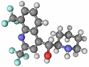
Background imageImages Dated 27th April 2011: Mefloquine malaria drug molecule

Mefloquine malaria drug molecule
Mefloquine, molecular model. This drug is used for the prevention and treatment of malaria. Atoms are represented as spheres and are colour-coded: carbon (grey), hydrogen (white)

Background imageImages Dated 27th April 2011: Trazadone antidepressant drug molecule

Trazadone antidepressant drug molecule
Trazadone, molecular model. This antidepressant drug is a serotonin antagonist and reuptake inhibitor (SARI). Atoms are represented as spheres and are colour-coded: carbon (grey), hydrogen (white)

Background imageImages Dated 27th April 2011: Modafinil stimulant drug molecule

Modafinil stimulant drug molecule
Modafinil, molecular model. This stimulant drug is used to treat narcolepsy. Atoms are represented as spheres and are colour-coded: carbon (grey), hydrogen (white), oxygen (red)

Background imageImages Dated 27th April 2011: Valsartan drug molecule

Valsartan drug molecule
Valsartan, molecular model. This drug is used to treat hypertension (high blood pressure) and congestive heart failure. Atoms are represented as spheres and are colour-coded: carbon (grey)

Background imageImages Dated 27th April 2011: Leflunomide arthritis drug molecule

Leflunomide arthritis drug molecule
Leflunomide, molecular model. This immunosuppressant and anti-inflammatory drug is used to treat rheumatoid and psoriatic arthritis

Background imageImages Dated 27th April 2011: Tubocurarine muscle relaxant molecule

Tubocurarine muscle relaxant molecule
Tubocurarine, molecular model. This muscle relaxant drug is obtained from the South American Chondrodendron tomentosum plant

Background imageImages Dated 27th April 2011: Phosgene chemical weapon molecule

Phosgene chemical weapon molecule
Phosgene chemical weapon, molecular model. Atoms are represented as spheres and are colour-coded: carbon (grey), chlorine (green) and oxygen (red)

Background imageImages Dated 27th April 2011: Phosphorus crystal structure

Phosphorus crystal structure, molecular model

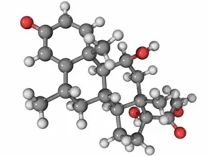
Background imageImages Dated 27th April 2011: Methylprednisolone drug molecule

Methylprednisolone drug molecule
Methylprednisolone, molecular model. This is a synthetic corticosteroid drug. Atoms are represented as spheres and are colour-coded: carbon (grey), hydrogen (white) and oxygen (red)

Background imageImages Dated 27th April 2011: Effexor antidepressant drug molecule

Effexor antidepressant drug molecule
Effexor. Molecular model of the selective serotonin reuptake inhibitor (SSRI) antidepressant drug venlafaxine. This drug is marketed as Effexor

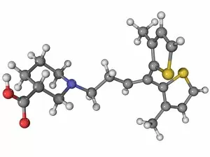
Background imageImages Dated 27th April 2011: Tiagabine epilepsy drug molecule

Tiagabine epilepsy drug molecule
Tiagabine, molecular model. This drug, marketed as Gabitril, is used to prevent seizures in epileptics. Atoms are represented as spheres and are colour-coded: carbon (grey), hydrogen (white)

Background imageImages Dated 27th April 2011: Phosgene chemical weapon molecule

Phosgene chemical weapon molecule
Phosgene chemical weapon, molecular model. Atoms are represented as spheres and are colour-coded: carbon (grey), chlorine (green) and oxygen (red)

Background imageImages Dated 27th April 2011: Kekulene hydrocarbon molecule

Kekulene hydrocarbon molecule, molecular model. Atoms are represented as spheres and are colour-coded: carbon (grey), hydrogen (white)

Background imageImages Dated 27th April 2011: Intestinal microvilli, TEM

Intestinal microvilli, TEM
Intestinal microvilli, coloured transmission electron micrograph (TEM). The microvilli are finger-like projections (seen here in circular cross-section)

Background imageImages Dated 27th April 2011: Peyers patches, SEM

Peyers patches, SEM
Peyers patches (red), coloured scanning electron micrograph (SEM). These regions of lymphoid tissue are found in the human intestine, usually in the ileum, the final section of the small intestine

Background imageImages Dated 27th April 2011: Intestinal villi, SEM

Intestinal villi, SEM
Intestinal villi, coloured scanning electron micrograph (SEM). The villi are finger-like projections that line the surface of the small intestine

Background imageImages Dated 27th April 2011: Intestinal villi cell loss, SEM

Intestinal villi cell loss, SEM
Intestinal villi cell loss, coloured scanning electron micrograph (SEM). Close-up of the tips of villi in the small intestine

Background imageImages Dated 27th April 2011: Intestinal villus cell loss, SEM

Intestinal villus cell loss, SEM
Intestinal villus cell loss, coloured scanning electron micrograph (SEM). Close-up of the tip of a villus in the small intestine that has shed a cell from its tip as part of the normal cell

Background imageImages Dated 27th April 2011: Intestinal villi cell loss, SEM

Intestinal villi cell loss, SEM
Intestinal villi cell loss, coloured scanning electron micrograph (SEM). Close-up of the tips of villi in the small intestine

Background imageImages Dated 27th April 2011: Peyers patches, SEM

Peyers patches, SEM
Peyers patches (green), coloured scanning electron micrograph (SEM). These regions of lymphoid tissue are found in the human intestine, usually in the ileum, the final section of the small intestine

Background imageImages Dated 27th April 2011: Intestinal villi cell loss, SEM

Intestinal villi cell loss, SEM
Intestinal villi cell loss, coloured scanning electron micrograph (SEM). Close-up of the tips of villi in the small intestine

Background imageImages Dated 27th April 2011: Intestinal microvilli, TEM

Intestinal microvilli, TEM
Intestinal microvilli, coloured transmission electron micrograph (TEM). The microvilli are finger-like projections (seen here in circular cross-section)

Background imageImages Dated 27th April 2011: Intestinal villi, SEM

Intestinal villi, SEM
Intestinal villi, coloured scanning electron micrograph (SEM). The villi are finger-like projections that line the surface of the small intestine


All Professionally Made to Order for Quick Shipping


-

Why Choose Us?

We are a leading provider of Art Prints and Photo Gifts since 2003, working in partnership with a range of Sporting Clubs, Charities, Museums and Picture Libraries. A large share of profits from any of their images will go directly towards supporting that charity or club. Our archive of images is carefully curated to bring you a wide range of subjects, including landscapes, wildlife, architecture, and more. We ship from our partner labs in the UK, USA, EU (Netherlands) and Australia.
+

How do I place an order?

Ordering is quick & easy - Just follow 5 Simple Steps:
  1. Find your image: Use our search box or browse our online photo Collections to find the image you want.

  2. Choose your print format: Select your desired product and add it to your cart.

  3. Enter your details: If you're a returning customer, simply enter your email address and password, and we'll fill in your billing and shipping address details. All personal details are held securely and are fully GDPR compliant. As standard, we remove all Personally Identifiable Information after 12 months.

  4. Pay for your purchase: We use state-of-the-art security for online shopping and do not have access to your card details.

  5. Sit back and relax: We'll email you confirmation of your order and when it's dispatched. Registered customers can also track orders in the 'My Account' area.

+

How do I pay for an order?

You can pay for your order with most of the major credit and debit cards, or PayPal. For added security, major financial institutions process payment details separately and securely on our behalf. We do not have access to your online payment card details. Online payments are preferred however we do take phone orders. For UK customers only we accept cheques issued against a UK bank.
+

Is my data safe?

We take data security very seriously. We do not have access to your full card details and all payments are requested over a fully secure connection. Additionally, we fully comply with current European and GDPR legislation, and automatically remove all personal data after at most 12 months (unless you have an account with us and have signed in recently). We also have a strict opt-in policy and would never sell your personal details. Your data is only used to fulfill your order promptly and efficiently.
+

Is wall art ready to hang?

For quick and easy installation all wall art, including framed prints, canvas prints and metal prints are supplied with a ready to hang solution on the back. Generally, saw tooth hangers are applied as they allow wall art to hang flush against the wall. The serrated edge of the hanger prevents the frame from shifting or tilting when hung.
+

Are the photo prints fade resistant?

Yes, we use archival quality photo paper photographic paper for vivid reproduction Prints are an accurate representations of the original artwork, which is preserved for artistic character and authenticity. We guarantee they match previews shown on our web site