Fraud Blocker Skip to main content

Home > Science Photo Library > Images Dated > 2006 > January > 4 Jan 2006

Images Dated 4th January 2006

Choose a picture from our Images Dated 4th January 2006 Collection for your Wall Art and Photo Gifts

32 items

We are proud to offer this selection in partnership with Science Photo Library

Background imageImages Dated 4th January 2006: Male shoot of cord moss

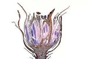
Male shoot of cord moss. Light micrograph of the top of a male shoot of the cord moss (Polytrichum commune). Antheridia (purple stain) are the male reproductive organs produce male gametes

Background imageImages Dated 4th January 2006: Liverwort spore case, light micrograph

Liverwort spore case, light micrograph
Liverwort spore case. Light micrograph of a vertical section through a spore case (sporophyte) from a liverwort (Pellia epiphylla)

Background imageImages Dated 4th January 2006: Gerbera flowers

Gerbera flowers (Gerbera sp.)

Background imageImages Dated 4th January 2006: Desk lamp

Desk lamp

Background imageImages Dated 4th January 2006: Dandelion (Taraxicum officinale)

Dandelion (Taraxicum officinale). Light micrograph of the flowerhead capitulum at the level of anthers of the multitude of flower florets, arranged in a fibonacci series

Background imageImages Dated 4th January 2006: Tropical storm Zeta, 2nd January 2006

Tropical storm Zeta, 2nd January 2006
Tropical storm Zeta over the Atlantic Ocean on 2nd January 2006. Zeta was the 27th named storm of the 2005 Atlantic hurricane season, extending the already record-breaking season yet further

Background imageImages Dated 4th January 2006: Starch grains in potato, light micrograph

Starch grains in potato, light micrograph
Starch grains in potato. Light micrograph of a potato (Solanum tuberosum) showing cells full of food starch grains

Background imageImages Dated 4th January 2006: Moss leaf cells, light micrograph

Moss leaf cells, light micrograph
Moss leaf cells. Polarised light micrograph of cells in a leaf of shining Hookeria moss (Hookeria luscens). Two whole cells (vertical) are seen at centre, surrounded by parts of other cells

Background imageImages Dated 4th January 2006: Plant stem trichomes, light micrograph

Plant stem trichomes, light micrograph
Plant stem trichomes. Polarised light micrograph of simple multicellular hairs (trichomes) on the surface (epidermis) of a stem of hedge woundwort (Stachys sylvatica)

Background imageImages Dated 4th January 2006: Rose stem, light micrograph

Rose stem, light micrograph
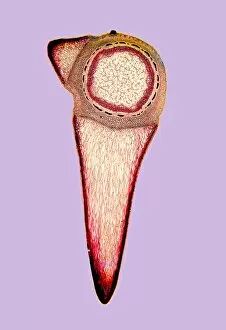
Rose stem. Light micrograph of a cross-section through the stem of a burnet rose (Rosa spinosisima), The two triangular positions are protective thorns

Background imageImages Dated 4th January 2006: Water-lily leaf stem, light micrograph

Water-lily leaf stem, light micrograph
Water-lily leaf stem. Light micrograph of a cross- section through the leaf stem (petiole) of a white water-lily (Nymphaea alba)

Background imageImages Dated 4th January 2006: Stem of a dracaena tree, micrograph

Stem of a dracaena tree, micrograph
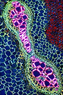
Stem of a Dracaena. Light micrograph of the stem of a Dracaena (Darcaena terminalis) of the Agavaceae (Monocotyledons), showing two vascular bundles containing fibres, found in the ground tissue

Background imageImages Dated 4th January 2006: Common horsetail stem, light micrograph

Common horsetail stem, light micrograph
Common horsetail stem. Light micrograph of a cross-section through the tip of the condensed stem of the common horsetail (Equisetum arvense), showing the central stem sourrounded by stem leaves

Background imageImages Dated 4th January 2006: Maple stem, light micrograph

Maple stem, light micrograph
Maple stem. Light micrograph of a cross-section of a stem from the field maple (Acer campestre). This shows the thick layer of cork (phellem) on the outside of the stem which insulates the stem

Background imageImages Dated 4th January 2006: Legume root nodules, light micrograph

Legume root nodules, light micrograph
Legume root nodules. Light micrograph of sections through two root nodules from a broad bean plant (Vicia faber). These root nodules are full of nitrogen-fixing bacteria

Background imageImages Dated 4th January 2006: Cucumber stem tissue, light micrograph

Cucumber stem tissue, light micrograph
Cucumber stem tissue. Light micrograph of a cross- section through the stem of a cucumber (Cucurbita sp.) showing a close up of the sieve cells (blue, two at centre left)

Background imageImages Dated 4th January 2006: Common horsetail stem, light micrograph

Common horsetail stem, light micrograph
Common horsetail stem. Light micrograph of a longitudinal section through the tip of the condensed stem of the giant horsetail (Equisetum telematia)

Background imageImages Dated 4th January 2006: Primary wood, light micrograph

Primary wood, light micrograph
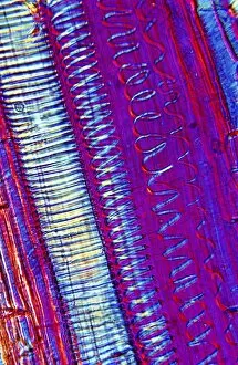
Primary wood of a tree. Light micrograph of a vertical section of the primary wood or xylem of a tree showing wood vessels with lignified annular, spiral and scalariform supporting thickenings

Background imageImages Dated 4th January 2006: Rhizome, light micrograph

Rhizome, light micrograph
Cross-section of a rhizome. Light micrograph of a cross-section through a rhizome of the bracken (Pterdium aquilinum) showing ground tissue in which there is imbedded a vascular strand called

Background imageImages Dated 4th January 2006: Tree xylem vessels, light micrograph

Tree xylem vessels, light micrograph
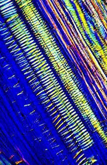
Tree xylem vessels. Polarised light micrograph of a vertical section through the primary wood vessels (xylem vessels) of a tree

Background imageImages Dated 4th January 2006: Leaf stem node, light micrograph

Leaf stem node, light micrograph
Leaf stem node. Light micrograph of a vertical section through a leaf stem node from a sycamore tree (Acer pseudoplatanus)

Background imageImages Dated 4th January 2006: Stem of the nootka cypress

Stem of the nootka cypress. Light micrograph of a cross-section of the stem of the conifer nootka cypress (Chamaecyparis nootkatensis)

Background imageImages Dated 4th January 2006: Broad bean root nodules, light micrograph

Broad bean root nodules, light micrograph
Broad bean root nodules. Light micrograph of the sections of the root nodules of the broad bean (Vicia faber) which are full of nitrogen-fixing bacteria

Background imageImages Dated 4th January 2006: Cucumber stem, light micrograph

Cucumber stem, light micrograph
Cucumber stem. Light micrograph of a cross-section through part of a vascular bundle from a cucumber (Cucurbita sp.). This vascular bundle is from the stem of the cucumber

Background imageImages Dated 4th January 2006: Wheat grain, light micrograph

Wheat grain, light micrograph
Wheat grain. Light micrograph of a vertical section through a grain of wheat (Triticum vulgare). This is the fruit of the plant, and includes the endosperm (larger area, pink)

Background imageImages Dated 4th January 2006: Double-seeded fruit, light micrograph

Double-seeded fruit, light micrograph
Double-seeded fruit. Light micrograph of a cross- section through a fruit with two seeds. This is the fruit of an Alexanders plant (Smyrnium olusatrum)

Background imageImages Dated 4th January 2006: Stellate leaf hairs, light micrograph

Stellate leaf hairs, light micrograph
Stellate leaf hairs. Polarised light micrograph of the epidermis of a leaf of the ornamental shrub Deutzia scabra, showing six of its stellate (star- shaped) hairs (trichomes). D

Background imageImages Dated 4th January 2006: Maize grain, light micrograph

Maize grain, light micrograph
Maize grain. Light micrograph of a vertical section through the grain of a maize plant (Zea mays). This is the fruit of the plant, and includes the endosperm (larger area, upper right, light pink)

Background imageImages Dated 4th January 2006: Leaf stomata, light micrograph

Leaf stomata, light micrograph
Leaf stomata. Light micrograph of a vertical section down through the surface of a leaf from a Scots pine tree (Pinus sylvestris)

Background imageImages Dated 4th January 2006: Wheat grain, light micrograph

Wheat grain, light micrograph
Wheat grain. Light micrograph with drak ground illumination of a vertical section through a grain of wheat (Triticum vulgare)

Background imageImages Dated 4th January 2006: Compound starch grains, light micrograph

Compound starch grains, light micrograph
Compound starch grains. Light micrograph of compound starch grains in the parenchyma cells of Phajus glandifolius, taken under polarised light


All Professionally Made to Order for Quick Shipping


-

Why Choose Us?

We are a leading provider of Art Prints and Photo Gifts since 2003, working in partnership with a range of Sporting Clubs, Charities, Museums and Picture Libraries. A large share of profits from any of their images will go directly towards supporting that charity or club. Our archive of images is carefully curated to bring you a wide range of subjects, including landscapes, wildlife, architecture, and more. We ship from our partner labs in the UK, USA, EU (Netherlands) and Australia.
+

How do I place an order?

Ordering is quick & easy - Just follow 5 Simple Steps:
  1. Find your image: Use our search box or browse our online photo Collections to find the image you want.

  2. Choose your print format: Select your desired product and add it to your cart.

  3. Enter your details: If you're a returning customer, simply enter your email address and password, and we'll fill in your billing and shipping address details. All personal details are held securely and are fully GDPR compliant. As standard, we remove all Personally Identifiable Information after 12 months.

  4. Pay for your purchase: We use state-of-the-art security for online shopping and do not have access to your card details.

  5. Sit back and relax: We'll email you confirmation of your order and when it's dispatched. Registered customers can also track orders in the 'My Account' area.

+

How do I pay for an order?

You can pay for your order with most of the major credit and debit cards, or PayPal. For added security, major financial institutions process payment details separately and securely on our behalf. We do not have access to your online payment card details. Online payments are preferred however we do take phone orders. For UK customers only we accept cheques issued against a UK bank.
+

Is my data safe?

We take data security very seriously. We do not have access to your full card details and all payments are requested over a fully secure connection. Additionally, we fully comply with current European and GDPR legislation, and automatically remove all personal data after at most 12 months (unless you have an account with us and have signed in recently). We also have a strict opt-in policy and would never sell your personal details. Your data is only used to fulfill your order promptly and efficiently.
+

Is wall art ready to hang?

For quick and easy installation all wall art, including framed prints, canvas prints and metal prints are supplied with a ready to hang solution on the back. Generally, saw tooth hangers are applied as they allow wall art to hang flush against the wall. The serrated edge of the hanger prevents the frame from shifting or tilting when hung.
+

Are the photo prints fade resistant?

Yes, we use archival quality photo paper photographic paper for vivid reproduction Prints are an accurate representations of the original artwork, which is preserved for artistic character and authenticity. We guarantee they match previews shown on our web site