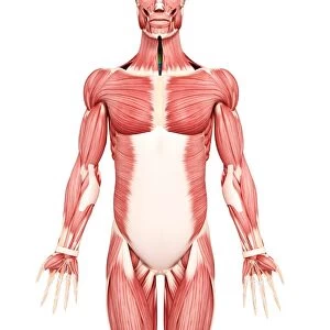
Premium Framed Print : Human foot musculature, artwork F007 / 3304


![]()
Framed Photos From Science Photo Library
Human foot musculature, artwork F007 / 3304
Human foot musculature, computer artwork
Science Photo Library features Science and Medical images including photos and illustrations
Media ID 9272535
© PIXOLOGICSTUDIO/SCIENCE PHOTO LIBRARY
Extensor Digitorum Brevis Extensor Digitorum Longus Extensor Hallucis Brevis Extensor Hallucis Longus Foot High Angle View Human Body Part Human Foot Human Leg Joint Limb Metatarsal Peroneus Brevis Phalanx Tarsal Tendon Tibia Tibialis Anterior Human Skeleton Musculature
14"x16" Premium Frame
Contemporary style Premium Wooden Frame with 8"x10" Print. Complete with 2" White Mat and 1.25" thick MDF frame. Printed on 260 gsm premium paper. Glazed with shatter proof UV coated acrylic glass. Backing is paper covered backing with rubber bumpers. Supplied ready to hang with a pre-installed sawtooth/wire hanger. Care Instructions: Spot clean with a damp cloth. Securely packaged in a clear plastic bag and envelope in a reinforced cardboard shipper
FSC Real Wood Frame and Double Mounted with White Conservation Mountboard - Professionally Made and Ready to Hang
Estimated Image Size (if not cropped) is 20.3cm x 25.4cm (8" x 10")
Estimated Product Size is 35.6cm x 40.6cm (14" x 16")
These are individually made so all sizes are approximate
Artwork printed orientated as per the preview above, with portrait (vertical) orientation to match the source image.
EDITORS COMMENTS
This print showcases the intricate musculature of the human foot, beautifully depicted through computer artwork. Against a pristine white background, this front view illustration offers a detailed glimpse into the biology and anatomy of a healthy adult foot. Every muscle, joint, and toe is meticulously portrayed, providing an invaluable resource for those interested in understanding the complexities of our lower limb. The image highlights various key components such as bones including tibia, tarsal, metatarsal, and phalanx structures that form the foundation of our feet. Additionally, tendons like tibialis anterior are prominently featured alongside other essential muscles like peroneus brevis, extensor digitorum brevis and longus as well as extensor hallucis brevis and longus. With its high angle perspective capturing every minute detail with precision and accuracy; this artwork serves as an indispensable tool for anatomical studies or medical research. The lifelike representation evokes awe at the marvels of human design while offering valuable insights into how our feet function on a biological level. Produced by PIXOLOGICSTUDIO/SCIENCE PHOTO LIBRARY – renowned for their commitment to scientific accuracy – this print exemplifies their dedication to delivering exceptional visual resources without compromising on quality or authenticity. A testament to both artistic skill and scientific knowledge combined seamlessly in one stunning composition.
MADE IN THE USA
Safe Shipping with 30 Day Money Back Guarantee
FREE PERSONALISATION*
We are proud to offer a range of customisation features including Personalised Captions, Color Filters and Picture Zoom Tools
SECURE PAYMENTS
We happily accept a wide range of payment options so you can pay for the things you need in the way that is most convenient for you
* Options may vary by product and licensing agreement. Zoomed Pictures can be adjusted in the Basket.