Premium Framed Print : Human foot musculature, artwork F007 / 2128


![]()
Framed Photos From Science Photo Library
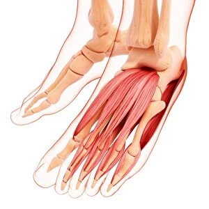
Human foot musculature, artwork F007 / 2128
Human foot musculature, computer artwork
Science Photo Library features Science and Medical images including photos and illustrations
Media ID 9267249
© PIXOLOGICSTUDIO/SCIENCE PHOTO LIBRARY
Calcaneus Extensor Digitorum Brevis Extensor Digitorum Longus Fibula Foot High Angle View Human Body Part Human Foot Joint Limb Metatarsal Phalanx Tarsal Tibia Human Skeleton Musculature Three Quarter View
16"x16" Premium Frame
Contemporary style Premium Wooden Frame with 10"x10" Print. Complete with 2" White Mat and 1.25" thick MDF frame. Printed on 260 gsm premium paper. Glazed with shatter proof UV coated acrylic glass. Backing is paper covered backing with rubber bumpers. Supplied ready to hang with a pre-installed sawtooth/wire hanger. Care Instructions: Spot clean with a damp cloth. Securely packaged in a clear plastic bag and envelope in a reinforced cardboard shipper
FSC Real Wood Frame and Double Mounted with White Conservation Mountboard - Professionally Made and Ready to Hang
Estimated Image Size (if not cropped) is 25.4cm x 25.4cm (10" x 10")
Estimated Product Size is 40.6cm x 40.6cm (16" x 16")
These are individually made so all sizes are approximate
Artwork printed orientated as per the preview above, with landscape (horizontal) or portrait (vertical) orientation to match the source image.
EDITORS COMMENTS
This print showcases the intricate musculature of the human foot, beautifully depicted through computer artwork. Against a pristine white background, this illustration captures the essence of adult biology, highlighting every muscle, joint, and toe with remarkable detail. The healthy anatomy of the foot is showcased in its normal state, providing a fascinating glimpse into this essential body part. From the fibula and tibia to the calcaneus and metatarsal bones, each component of the foot's structure is meticulously portrayed. This high-angle view offers a comprehensive understanding of how these elements work together to support our daily movements. The artist has masterfully represented not only the external features but also delves into internal details such as tendons like extensor digitorum brevis and extensor digitorum longus. This comprehensive representation adds depth to our comprehension of this complex anatomical region. With its three-quarter perspective and lifelike rendering, this artwork seamlessly combines scientific accuracy with artistic flair. It serves as an invaluable resource for students studying human anatomy or anyone seeking a deeper appreciation for our incredible skeletal system. This awe-inspiring piece by PIXOLOGICSTUDIO/SCIENCE PHOTO LIBRARY is more than just an image; it embodies both scientific knowledge and creative expression at their finest.
MADE IN THE USA
Safe Shipping with 30 Day Money Back Guarantee
FREE PERSONALISATION*
We are proud to offer a range of customisation features including Personalised Captions, Color Filters and Picture Zoom Tools
SECURE PAYMENTS
We happily accept a wide range of payment options so you can pay for the things you need in the way that is most convenient for you
* Options may vary by product and licensing agreement. Zoomed Pictures can be adjusted in the Basket.