Jigsaw Puzzle : Osteoporosis, artwork


![]()
Jigsaw Puzzles From Science Photo Library
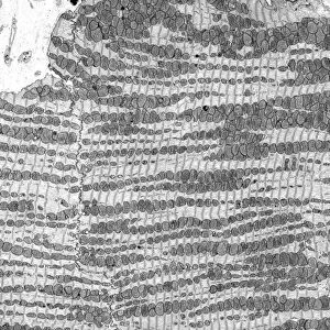
Osteoporosis, artwork
Osteoporosis. Artwork of vertical sections comparing the upper parts of a healthy femur (left) and one with reduced bone mass (right). The head of the femur (which articulates with the pelvis in the hip joint) is at left in each artwork. The loss of bone mass at right could be caused by a condition such as osteoporosis. In this condition, loss of calcium from the interior of bones leaves them brittle and prone to fracture. Broken hips due to falls occur in elderly women with this condition, which affects women more than men
Science Photo Library features Science and Medical images including photos and illustrations
Media ID 6325409
© HENNING DALHOFF / SCIENCE PHOTO LIBRARY
Brittle Comparing Comparison Femur Fragile Joint Osteology Osteoporosis Osteoporotic Reduction Sections Shaft Abnormal Section Sectioned Unhealthy
Jigsaw Puzzle (520 Pieces)
Discover the intricacies of the human body with our captivating Osteoporosis jigsaw puzzle from Media Storehouse. Featuring an enlightening artwork by Science Photo Library, this puzzle invites you to explore the differences between a healthy femur and one affected by osteoporosis. Ideal for both educational and therapeutic purposes, this puzzle provides a unique way to learn about bone health while engaging your mind and enhancing focus and concentration. Bring this fascinating puzzle into your home and embark on a journey of discovery, piece by piece.
Made in the USA, 520-piece puzzles measure 16" x 20" (40.6 x 50.8 cm). Every puzzle is meticulously printed on glossy photo paper, which has a strong 1.33 mm thickness. Delivered in a black storage cardboard box, these puzzles are both stylish and practical. (Note: puzzles contain small parts and are not suitable for children under 3 years of age.)
Jigsaw Puzzles are an ideal gift for any occasion
Estimated Product Size is 50.8cm x 40.5cm (20" x 15.9")
These are individually made so all sizes are approximate
Artwork printed orientated as per the preview above, with landscape (horizontal) or portrait (vertical) orientation to match the source image.
EDITORS COMMENTS
This artwork, titled "Osteoporosis" offers a visual comparison between the upper parts of a healthy femur and one with reduced bone mass. The print showcases vertical sections of both femurs side by side, highlighting the stark contrast in their composition. On the left, we see the robust structure of a normal femur, while on the right, we witness the detrimental effects of reduced bone mass caused by conditions like osteoporosis. The focus is primarily on the head of each femur as it articulates with the pelvis in the hip joint. This intricate joint plays a crucial role in our mobility and overall well-being. However, when calcium loss occurs within bones due to osteoporosis, they become brittle and susceptible to fractures. This condition predominantly affects women more than men and often leads to broken hips resulting from falls among elderly individuals. Through this thought-provoking illustration, viewers gain insight into how osteoporosis can impact our skeletal system's health and functionality. It serves as an educational tool for medical professionals studying anatomy or anyone interested in understanding this prevalent bone disease better. Created by Science Photo Library's talented artists using meticulous anatomical details combined with artistic flair, this print beautifully captures both scientific accuracy and aesthetic appeal.
MADE IN THE USA
Safe Shipping with 30 Day Money Back Guarantee
FREE PERSONALISATION*
We are proud to offer a range of customisation features including Personalised Captions, Color Filters and Picture Zoom Tools
SECURE PAYMENTS
We happily accept a wide range of payment options so you can pay for the things you need in the way that is most convenient for you
* Options may vary by product and licensing agreement. Zoomed Pictures can be adjusted in the Basket.