Fraud Blocker Skip to main content

Vertebra Collection (page 2)

"Unveiling the Backbone of Life: Exploring the Fascinating World of Vertebrae" A glimpse into history reveals a horse's skull

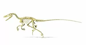
Background imageVertebra Collection: 3D rendering of a Velociraptor dinosaur skeleton

3D rendering of a Velociraptor dinosaur skeleton, side view

Background imageVertebra Collection: 3D rendering of a Diplodocus dinosaur skeleton

3D rendering of a Diplodocus dinosaur skeleton, side view. Diplodocus was a giant herbivorous dinosaur of the late Jurassic period

Background imageVertebra Collection: Spinosaurus dinosaur skeleton

Spinosaurus dinosaur skeleton on textured background

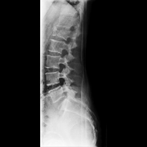
Background imageVertebra Collection: Fractured vertebra, X-ray C017 / 7586

Fractured vertebra, X-ray C017 / 7586
Fractured vertebra. X-ray of the lower spine of a 50 year old male with a fracture to the L1 lumbar vertebra (thord block from top)

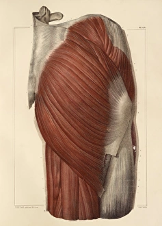
Background imageVertebra Collection: Pelvic-femoral muscles, 1831 artwork

Pelvic-femoral muscles, 1831 artwork
Pelvic-femoral muscles, superficial layer. Part of the spine at top left. This anatomical artwork is plate 124 from volume 2 (1831) of Traite complet de l anatomie de l homme (1831-1854)

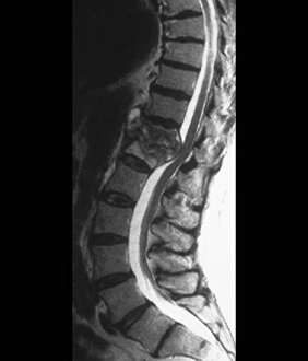
Background imageVertebra Collection: Tuberculosis of the spine, MRI scan

Tuberculosis of the spine, MRI scan
Tuberculosis of the spine. Magnetic resonance imaging (MRI) scan of a sagittal section through the spine of a 74-year-old patient with tuberculosis (TB) of the spine, or Potts disease

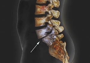
Background imageVertebra Collection: Spondylolisthesis, 3D CT scan

Spondylolisthesis, 3D CT scan
Spondylolisthesis. Coloured 3D computed tomography (CT) scan of a section through the lumbar (lower back) spine of a 39-year-old patient with spondylolisthesis

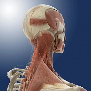
Background imageVertebra Collection: Neck muscles and nerves, artwork

Neck muscles and nerves, artwork
Neck and upper back muscles (pink) and nerves (yellow), computer artwork. The large opaque muscle covering most of the upper back is the trapezius

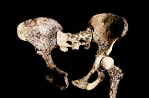
Background imageVertebra Collection: Australopithecus africanus pelvis, STS-14 C015 / 6919

Australopithecus africanus pelvis, STS-14 C015 / 6919
Australopithecus africanus pelvis (STS-14). This fossil specimen was discovered in 1947, in Sterkfontein, South Africa. The entire specimen consists of the pelvis, part of the vertebral column

Background imageVertebra Collection: Coccyx pain, artwork

Coccyx pain, artwork
Coccyx (tailbone) pain, computer artwork

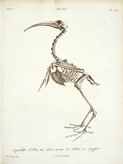
Background imageVertebra Collection: Threskiornis aethiopicus, sacred ibis skeleton

Threskiornis aethiopicus, sacred ibis skeleton
Plate 52 from a paper published by Georges Cuvier in 1804

Background imageVertebra Collection: Torso bones

Torso bones. Computer artwork of the bones of the human torso. The backbone (running vertically down the centre) is a flexible column of 26 bones (vertebrae)

Background imageVertebra Collection: Normal lumbar spine, X-ray

Normal lumbar spine, X-ray
Normal lumbar spine. Coloured frontal X-ray showing bones of the lower (lumbar) spine (centre, purple). The bones of the spine (block-shaped) are called vertebrae

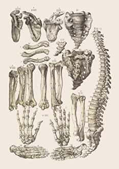
Background imageVertebra Collection: Bones of the human skeleton

Bones of the human skeleton
Bones of the skeleton. Artwork from Juan Valverde de Amuscos textbook Anatomia del Corpo Humano (Anatomy of the Human Body), published 1560. Fig. I is a view of the spine, from the left side. Figs

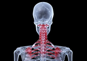
Background imageVertebra Collection: Neck and shoulder pain, X-ray artwork

Neck and shoulder pain, X-ray artwork
Neck and shoulder pain. Computer X-ray artwork of the back view of a human skeletons shoulders, neck and skull. The red area represents neck pain (cervicalgia) and shoulder pain

Background imageVertebra Collection: Normal female pelvis, X-ray

Normal female pelvis, X-ray
Normal female pelvis, coloured frontal X-ray. The thigh (femur) bones of the leg are seen at bottom left and right, and the base of the spine is at top

Background imageVertebra Collection: Skeleton lying down, artwork

Skeleton lying down, artwork
Skeleton lying down. Computer artwork showing the bones of the head, neck and upper torso of a human skeleton that is lying down

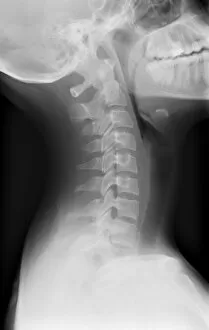
Background imageVertebra Collection: Healthy spine of the neck, X-ray

Healthy spine of the neck, X-ray
X-ray (side view) of the neck of an adult patient, showing healthy cervical vertebrae (spine bones) of the neck. The skull (at top) is tilted back, with the shoulders at lower frame

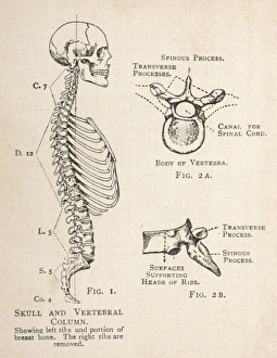
Background imageVertebra Collection: Diagram of human skull and vertebral column

Diagram of human skull and vertebral column
A diagram of the human skull and vertebral column, showing the left ribs and a portion of the breastbone, as well as a single vertebra from two angles

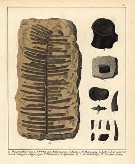
Background imageVertebra Collection: Fossil fern, Nothosaurus tooth and vertebra

Fossil fern, Nothosaurus tooth and vertebra, Dracorex tooth, and fossil fish scales.. Handcolored lithograph from Dr. F.A

Background imageVertebra Collection: Neck vertebra anatomy engraving 1866

Neck vertebra anatomy engraving 1866
Atlas d anatomie descriptive du corps humain C. Bonamy - Paul Broca Victor Masson et Fils Paris 1866

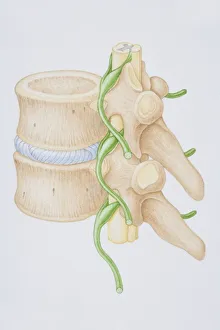
Background imageVertebra Collection: Section diagram depicting the human vertebral column, spinal nerve

Section diagram depicting the human vertebral column, spinal nerve, spinal cord and vertebra of the delicate spina

Background imageVertebra Collection: Digital illustration of lumbar puncture using spinal needle inserted into lumbar vertebrae

Digital illustration of lumbar puncture using spinal needle inserted into lumbar vertebrae and dura mater

Background imageVertebra Collection: Digital illustration Spina Bifida, Meningocele, where protective covering around spinal cord

Digital illustration Spina Bifida, Meningocele, where protective covering around spinal cord protrudes through malformed vertebra to form sac filled with cerebrospinal fluid

Background imageVertebra Collection: Digital illustration of head in profile showing cross section of brain, neck vertebra and spine

Digital illustration of head in profile showing cross section of brain, neck vertebra and spine

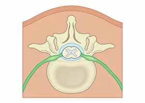
Background imageVertebra Collection: Cross section digital illustration of spinal cord and nerves surrounded by vertebra

Cross section digital illustration of spinal cord and nerves surrounded by vertebra

Background imageVertebra Collection: Cross section biomedical illustration of lumber microdisectomy before surgery

Cross section biomedical illustration of lumber microdisectomy before surgery

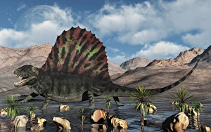
Background imageVertebra Collection: Dimetrodon reptile from the Permian period

Dimetrodon reptile from the Permian period
Dimetrodon was a carnivorous mammal-like reptile which lived during the Permian period of North America and Europe

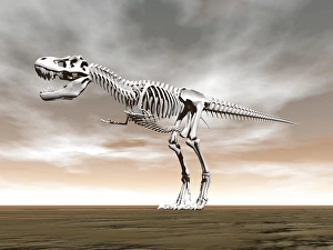
Background imageVertebra Collection: Tyrannosaurus rex skeleton

Tyrannosaurus rex skeleton

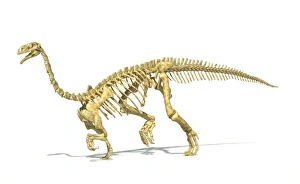
Background imageVertebra Collection: 3D rendering of a Plateosaurus dinosaur skeleton

3D rendering of a Plateosaurus dinosaur skeleton, perspective view

Background imageVertebra Collection: 3D rendering of an Iguanodon dinosaur skeleton

3D rendering of an Iguanodon dinosaur skeleton, perspective view

Background imageVertebra Collection: Dimetrodon was an extinct genus of synapsid from th Early Permian period

Dimetrodon was an extinct genus of synapsid from th Early Permian period
Dimetrodon was an extinct genus of synapsid that lived during the Early Permian, around 295a'272 million years ago

Background imageVertebra Collection: A sail-backed Dimetrodon from Earths Permian period of time

A sail-backed Dimetrodon from Earths Permian period of time

Background imageVertebra Collection: 3D rendering of a Dimetrodon dinosaur

3D rendering of a Dimetrodon dinosaur, side view

Background imageVertebra Collection: Allosaurus dinosaur skeleton

Allosaurus dinosaur skeleton on textured background

Background imageVertebra Collection: 3D rendering of a Pachycephalosaurus dinosaur skeleton

3D rendering of a Pachycephalosaurus dinosaur skeleton, perspective view

Background imageVertebra Collection: Comparison of Tyrannosaurus Rex standing next to its fossil skeleton

Comparison of Tyrannosaurus Rex standing next to its fossil skeleton

Background imageVertebra Collection: 3D rendering of an Allosaurus dinosaur skeleton

3D rendering of an Allosaurus dinosaur skeleton, side view. Allosaurs were large carnivorous reptiles that lived during the late Jurassic period

Background imageVertebra Collection: A group of sail-backed Dimetrodons during Earths Permian period

A group of sail-backed Dimetrodons during Earths Permian period
A group of sail-backed carnivorous Dimetrodons during Earths Permian period

Background imageVertebra Collection: 3D rendering of a Giganotosaurus dinosaur skeleton

3D rendering of a Giganotosaurus dinosaur skeleton, perspective view

Background imageVertebra Collection: An Ankylosaurus dinosaur with full skeleton superimposed

An Ankylosaurus dinosaur with full skeleton superimposed
3D rendering of an Ankylosaurus dinosaur with full skeleton superimposed, side view. This armored dinosaur lived in the early Mesozoic era

Background imageVertebra Collection: Sail-backed Dimetrodons, alive during Earths Permian period of time

Sail-backed Dimetrodons, alive during Earths Permian period of time

Background imageVertebra Collection: Ceratosaurus dinosaur skeleton

Ceratosaurus dinosaur skeleton on textured background

Background imageVertebra Collection: Giganotosaurus dinosaur skeleton

Giganotosaurus dinosaur skeleton on textured background

Background imageVertebra Collection: Diapsid - Megalania: Vertebra

Diapsid - Megalania: Vertebra

Background imageVertebra Collection: Diplodocus: Tail vertebra

Diplodocus: Tail vertebra

Background imageVertebra Collection: Cetiosaurus: Vertebra

Cetiosaurus: Vertebra


All Professionally Made to Order for Quick Shipping

"Unveiling the Backbone of Life: Exploring the Fascinating World of Vertebrae" A glimpse into history reveals a horse's skull, showcasing the intricate vertebrae that form its backbone. Delving deeper, a diagram illustrates the human brain and spinal column, highlighting how vital vertebrae are to our nervous system. With a camera in hand, an individual captures an X-ray image, unveiling the hidden secrets within vertebral structures. Journeying back in time, we encounter a Liopleurodon vertebra—a relic from prehistoric oceans—reminding us of ancient creatures' remarkable skeletal systems. Witnessing neck vertebrae extended through an X-ray image showcases their flexibility and adaptability for various movements. An artistically colored X-ray depicts lumbar vertebrae—the foundation of our spine—revealing their strength and interconnectedness. Examining a normal neck through an X-ray unveils its structural integrity—an essential component for supporting our head and facilitating movement. Immerse yourself in paleontological wonders with a 3D rendering of an Ankylosaurus dinosaur skeleton, emphasizing its robust vertebral framework. Peering inside another 3D rendering—this time featuring a Tyrannosaurus Rex dinosaur skeleton—we marvel at how these extinct giants relied on their powerful spines for survival. Unearthed from centuries past is an illustration depicting the nervous system as understood during the 18th century—a testament to humanity's evolving knowledge about vertebral significance over time. In modern times, workers collaborate to repair damaged spines—an inspiring reminder of medical advancements aimed at restoring mobility and quality of life for individuals facing spinal issues (Spine with workers; spine repair F007 / 9907).