Fraud Blocker Skip to main content

Vertebra Collection (page 7)

"Unveiling the Backbone of Life: Exploring the Fascinating World of Vertebrae" A glimpse into history reveals a horse's skull

Background imageVertebra Collection: Neck vertebrae flexed, X-ray

Neck vertebrae flexed, X-ray
Bending of the neck. Coloured X-ray of a side view of the neck of a man showing flexion of healthy cervical vertebrae (bones)

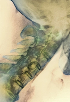
Background imageVertebra Collection: Neck vertebrae extended, X-ray

Neck vertebrae extended, X-ray
Bending of the neck. Coloured X-ray of a side view of the neck of a man showing extension of healthy cervical vertebrae (bones)

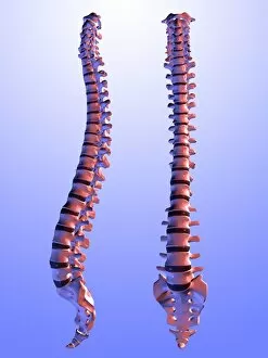
Background imageVertebra Collection: Two views of the human spine

Two views of the human spine. This computer artwork shows a side (lateral) view (left) and an front (anterior) view (right)

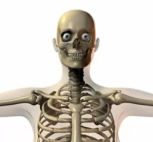
Background imageVertebra Collection: Upper body bones

Upper body bones, computer artwork. The skull (top) consists of the cranium, which encloses the brain and the jaw (mandible)

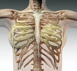
Background imageVertebra Collection: Ribcage

Ribcage and upper body, computer artwork. The spine, which consists of blocks of bone (vertebrae) separated by spongy intervertebral discs, runs down centre

Background imageVertebra Collection: Pelvis

Pelvis. Computer artwork showing the bones of a healthy pelvis. At top is the spine, which consists of blocks of bone (vertebrae) separated by spongy intervertebral disks

Background imageVertebra Collection: Skull

Skull. Computer artwork of the bones and musculature of a human head. The skull consists of the cranium, which encloses the brain, and the jaw. Embedded in the jaw are the teeth

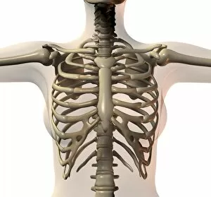
Background imageVertebra Collection: Female ribcage

Female ribcage and upper body, computer artwork. The spine, which consists of blocks of bone (vertebrae) separated by spongy intervertebral discs, runs down centre

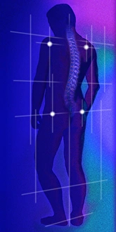
Background imageVertebra Collection: Computer illustratoion of a healthy human spine

Computer illustratoion of a healthy human spine
Spine. Computer illustration showing a spine highlighted against a silhouetted male human figure seen from the back. A grid superimposed on the silhouette

Background imageVertebra Collection: Spine

Spine. Computer artwork of a healthy spine. At top is the skull and at bottom are the bones of the pelvis. The spine is a column of 33 cylindrical bones called vertebrae

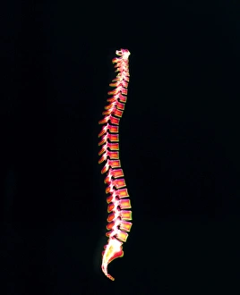
Background imageVertebra Collection: Spine

Spine. Computer artwork of a healthy spine seen from the side. At top is the skull and at bottom are the bones of the pelvis

Background imageVertebra Collection: Coloured X-ray of a healthy human spine

Coloured X-ray of a healthy human spine
Spine. Coloured X-ray showing a healthy human spine seen from the side. The front of the spine is at right. The spine is made of a column of 33 cylindrical bones called vertebrae which support

Background imageVertebra Collection: Healthy neck bones, X-ray

Healthy neck bones, X-ray
Healthy neck bones, coloured X-ray. The neck contains seven vertebrae, the blocky bones that make up the spinal column (down centre)

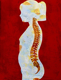
Background imageVertebra Collection: Artwork of spine on profile of a young girl

Artwork of spine on profile of a young girl

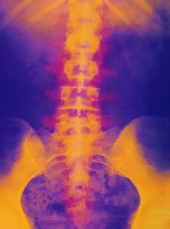
Background imageVertebra Collection: Coloured X-ray of normal lumbar spine (lower back)

Coloured X-ray of normal lumbar spine (lower back)
Lumbar spine and pelvis. Computer-enhanced X-ray of the healthy lumbar spine (lower back) and pelvis of a human. At top can be seen the lower ribs of the ribcage

Background imageVertebra Collection: Coloured X-ray of normal lower spine and pelvis

Coloured X-ray of normal lower spine and pelvis
Lower spine and pelvis. Coloured X-ray of healthy human lumbar spine (lower back) and pelvic girdle. At top can be seen the 11th and 12th floating ribs on either side of the thoracic vertebrae

Background imageVertebra Collection: Artwork of cervical vertebra from human spine

Artwork of cervical vertebra from human spine
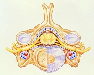
Cervical vertebra with spinal cord. Illustration of a bone from the human spine, showing blood vessels (red and blue), the spinal cord (yellow, centre), nerves (yellow) and cartilage (pale blue)

Background imageVertebra Collection: Coloured X-ray of a normal neck in profile

Coloured X-ray of a normal neck in profile
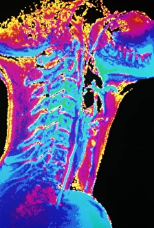
Human neck. Coloured X-ray of a normal human neck in profile. The seven cervical vertebrae (spinal bones) are clearly identifiable in the centre

Background imageVertebra Collection: Spinal column

Spinal column. Historical artwork of the bones of the human spine and pelvis, seen from the front. The backbone (vertical)

Background imageVertebra Collection: Spinal vertebra

Spinal vertebra. Historical artwork of a vertebra (spinal bone) seen from the side. The rear of the spine is at right. This is a lumbar vertebra, one from the lower back

Background imageVertebra Collection: Spinal vertebrae

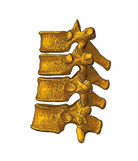
Spinal vertebrae. Historical artwork of vertebrae (spinal bones) seen from the side. The rear of the spine is at right. Four vertebrae are seen here

Background imageVertebra Collection: Spinal bones and ligaments

Spinal bones and ligaments. Historical anatomical artwork of thoracic (upper) spinal bones (yellow, vertebrae) and ligaments (white)

Background imageVertebra Collection: Lower back pain, X-ray artwork

Lower back pain, X-ray artwork
Back pain. Computer X-ray artwork of the rear view of the lower back of a human skeleton. The red area represents lower back pain (lumbago)

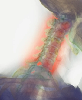
Background imageVertebra Collection: Neck pain, X-ray artwork

Neck pain, X-ray artwork
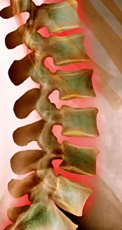
Neck pain. Computer X-ray artwork of a lateral view of a the bones of the neck. The front of the neck is at left. The red area represents neck pain (cervicalgia)

Background imageVertebra Collection: Back pain, X-ray artwork

Back pain, X-ray artwork
Back pain. Computer X-ray artwork of the rear view of a human skeleton. The red area represents back pain (dorsalgia). Back pain usually originates from the muscles, nerves, bones
Background imageVertebra Collection: Lower back pain, X-ray

Lower back pain, X-ray
Lower back pain. Conceptual coloured X-ray of the bones of the lower back glowing red in pain. Lower back pain (lumbago) is a very common complaint

Background imageVertebra Collection: Osteoporosis of the back, X-ray

Osteoporosis of the back, X-ray
Osteoporosis of the back, coloured X-ray. Osteoporosis is a condition in which bone density is reduced, weakening the bones and making them susceptible to fractures

Background imageVertebra Collection: Pinned curved spine, X-ray

Pinned curved spine, X-ray
Pinned curved spine. Coloured X-ray of bones of the spine and pelvis of a 49 year old woman showing two screws (blue) pinning the lower lumbar vertebra (L5) to the sacrum of the pelvis

Background imageVertebra Collection: Scoliosis spine deformity, X-ray

Scoliosis spine deformity, X-ray
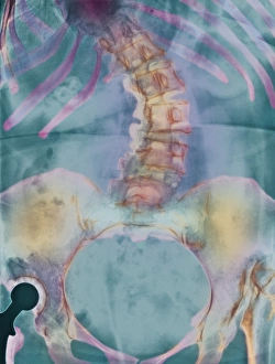
Scoliosis sideways deformity of the spine of a woman, coloured X-ray. The spine (upper centre) is severely bent. The deformity affected the patients posture

Background imageVertebra Collection: Osteoporotic spine, X-ray

Osteoporotic spine, X-ray
Osteoporotic spine, coloured X-ray. The spine is seen in side view, with the front of the body at right. It is seen to be curved, bowing out from top right to centre left then back right again

Background imageVertebra Collection: Osteoporosis of the spine, CT scan

Osteoporosis of the spine, CT scan
Osteoporosis of the spine. Coloured 3D computed tomography (CT) scan of the twisted lumbar vertebrae of a woman with osteoporosis

Background imageVertebra Collection: Spine degeneration, MRI scan

Spine degeneration, MRI scan
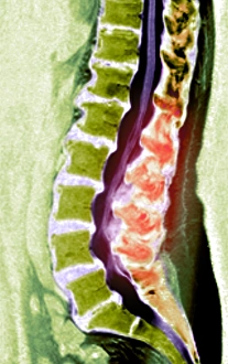
Spine degeneration. Coloured magnetic resonance imaging (MRI) scan of the lumbar (lower) region of a patients back showing spinal degeneration. The base of the spine is at lower right

Background imageVertebra Collection: Pinned vertebrae, X-ray

Pinned vertebrae, X-ray
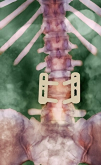
Pinned vertebrae. Coloured X-ray of pins (beige) joining two of the vertebrae (pink) of a 29 year old woman together to immobilise them. Vertebrae are the box-like bones that make up the spine

Background imageVertebra Collection: Slipped disc, CT scan

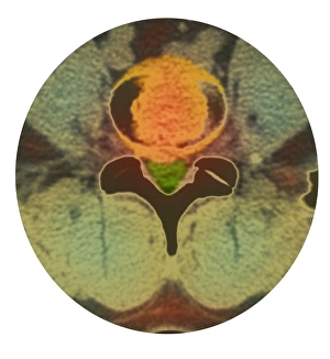
Slipped disc, CT scan
Slipped disc. Coloured computed tomography (CT) scan of an axial section through an inter- vertebral disc (orange) showing herniation

Background imageVertebra Collection: Spinal osteoporosis, X-ray

Spinal osteoporosis, X-ray
Spinal osteoporosis. Coloured X-ray of the lumbar region of the spine of an elderly female patient with osteoporosis. The bones have lost their density and have become porous and fragile

Background imageVertebra Collection: Osteoporosis of spine, X-ray

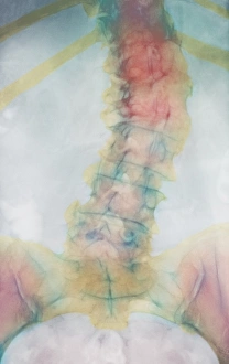
Osteoporosis of spine, X-ray
Osteoporosis. Coloured X-ray of a rear view of the lumbar (lower) spine of an elderly woman showing scoliosis (sideways curvature) of the spine caused by osteoporosis

Background imageVertebra Collection: Coloured gamma scan of osteoporosis in vertebra

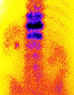
Coloured gamma scan of osteoporosis in vertebra
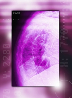
Osteoporosis. Coloured gamma scan (scintigram) of a spine showing vertebral damage (dark) due to osteoporosis. Osteoporosis is a lowering of bone density causing bones to become brittle

Background imageVertebra Collection: Computer artwork of osteoporosis affecting bone

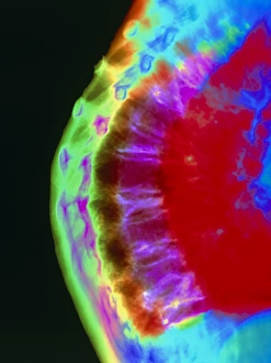
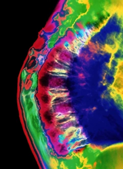
Computer artwork of osteoporosis affecting bone
Osteoporosis. Computer artwork of a Scanning Electron Micrograph showing the effect of osteoporosis on bone tissue. This section through one of the vertebrae of the backbone shows the cancellous

Background imageVertebra Collection: Coloured X-ray of curved spine in osteoporosis

Coloured X-ray of curved spine in osteoporosis
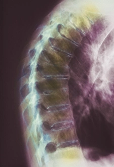
Curved spine in osteoporosis. Computer-enhanced X- ray of the curved and deformed thoracic spine of a woman suffering from osteoporosis

Background imageVertebra Collection: Spinal osteoporosis

Spinal osteoporosis
Curved spine in osteoporosis. Computer-enhanced X-ray of the curved and deformed thoracic spine of a woman suffering from osteoporosis

Background imageVertebra Collection: Osteoarthritis of neck, X-ray

Osteoarthritis of neck, X-ray
Osteoarthritis of the neck. Coloured X-ray of a side view of the neck of an elderly man, showing degenerative osteoarthritis of the cervical bones of the neck. Also known as cervical spondylosis

Background imageVertebra Collection: Ankylosing spondylitis X-ray

Ankylosing spondylitis X-ray
Ankylosing spondylitis. Coloured X-ray of the spine of a patient with ankylosing spondylitis, a form of arthritis. This is the lumbar region in the lower back

Background imageVertebra Collection: Bony spurs in neck, X-ray

Bony spurs in neck, X-ray
Bony spurs in neck, coloured X-ray. Bony spurs (osteophytes, left) grow from the vertebrae as a normal part of the aging process

Background imageVertebra Collection: Degenerative disc disease, 3D CT scan

Degenerative disc disease, 3D CT scan
Degenerative disc disease. Coloured 3D computed tomography (CT) scan of the lumbosacral spine of a 74 year old patient with degenerative disc disease

Background imageVertebra Collection: Healthy kidneys, artwork

Healthy kidneys, artwork


All Professionally Made to Order for Quick Shipping

"Unveiling the Backbone of Life: Exploring the Fascinating World of Vertebrae" A glimpse into history reveals a horse's skull, showcasing the intricate vertebrae that form its backbone. Delving deeper, a diagram illustrates the human brain and spinal column, highlighting how vital vertebrae are to our nervous system. With a camera in hand, an individual captures an X-ray image, unveiling the hidden secrets within vertebral structures. Journeying back in time, we encounter a Liopleurodon vertebra—a relic from prehistoric oceans—reminding us of ancient creatures' remarkable skeletal systems. Witnessing neck vertebrae extended through an X-ray image showcases their flexibility and adaptability for various movements. An artistically colored X-ray depicts lumbar vertebrae—the foundation of our spine—revealing their strength and interconnectedness. Examining a normal neck through an X-ray unveils its structural integrity—an essential component for supporting our head and facilitating movement. Immerse yourself in paleontological wonders with a 3D rendering of an Ankylosaurus dinosaur skeleton, emphasizing its robust vertebral framework. Peering inside another 3D rendering—this time featuring a Tyrannosaurus Rex dinosaur skeleton—we marvel at how these extinct giants relied on their powerful spines for survival. Unearthed from centuries past is an illustration depicting the nervous system as understood during the 18th century—a testament to humanity's evolving knowledge about vertebral significance over time. In modern times, workers collaborate to repair damaged spines—an inspiring reminder of medical advancements aimed at restoring mobility and quality of life for individuals facing spinal issues (Spine with workers; spine repair F007 / 9907).