Fraud Blocker Skip to main content

Reproductive Collection (page 15)

"Exploring the Intricate World of Reproduction: From Dinosaurs to Flowers and Beyond" In the ancient world

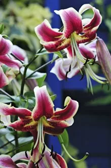
Background imageReproductive Collection: Lily (Lilium Scheherazade )

Lily (Lilium Scheherazade )
Family: Liliaceae Genus: lilium Cultivar/hybrid Scheherazade Oriental trumpet lily, Dorset, UK August 2010

Background imageReproductive Collection: Magpie aquilegia (Aquilegia sp. )

Magpie aquilegia (Aquilegia sp. )
Magpie aquilegia in flower. Dorset, UK June 2010

Background imageReproductive Collection: Pelargonium Black Prince

Pelargonium Black Prince Dorset, UK July 2010

Background imageReproductive Collection: Nectaroscordum scopulorum

Nectaroscordum scopulorum
Nectaruscordum scopulorum coming into bloom. Dorset, UK July 2010

Background imageReproductive Collection: Penguins mating, artwork

Penguins mating, artwork. The penguins reproductive organs are shown here, with the male on top and the female on the bottom

Background imageReproductive Collection: Seychelles sunbird nest

Seychelles sunbird nest. This nest, built by the Seychelles sunbird (Cinnyris dussumieri), is usually made from grass and moss, and bound with spiders webs. Numerous feathers are also seen here

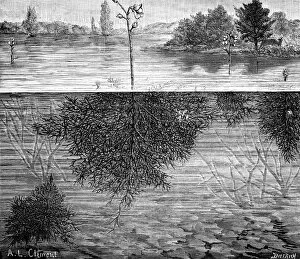
Background imageReproductive Collection: Bladderwort in a lake

Bladderwort in a lake. The bladderworts (Utricularia) are carnivorous plants. The underwater bladders trap small aquatic animals, such as water fleas or even small fish fry and tadpoles

Background imageReproductive Collection: Oviraptors dinosaurs nesting

Oviraptors dinosaurs nesting
Oviraptor dinosaurs nesting. Artwork of two Oviraptor dinosaurs camouflaged on their nest. The fossils of this dinosaur were found in Mongolia, dating to around 75 million years ago

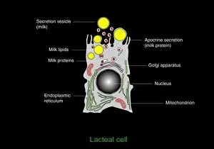
Background imageReproductive Collection: Milk-producing cell, artwork

Milk-producing cell, artwork
Milk-producing cell. Artwork of the anatomical structure of a lacteal (milk-producing) cell. Normal cell organelles include the nucleus (black, centre), the golgi apparatus (white)

Background imageReproductive Collection: Rare smalleye stingra

Rare smalleye stingra
Sperm cell. Artwork of a spermatozoon, a human male sex cell. At left is the head, containing the genetic material in the nucleus

Background imageReproductive Collection: Peony pollen grains, SEM

Peony pollen grains, SEM
Peony (Paeonia sp.) pollen grains, coloured scanning electron micrograph (SEM). Pollen grains are reproductive structures produced by the male parts of flowering plants

Background imageReproductive Collection: Milk-producing cell, diagram

Milk-producing cell, diagram
Milk-producing cell. Diagram of the anatomical structure of a lacteal (milk-producing) cell. Normal cell organelles include the nucleus (black, centre), the golgi apparatus (white)

Background imageReproductive Collection: Dandelion seed, SEM

Dandelion seed, SEM
Dandelion (Taraxacum officinale) seed, coloured scanning electron micrograph (SEM)

Background imageReproductive Collection: Pentaceratops dinosaurs mating

Pentaceratops dinosaurs mating. Artwork of male (right) and female (left) Pentaceratops dinosaurs mating. The name of this herbivorous dinosaur refers to the five horns on its head

Background imageReproductive Collection: Human fertilisation, artwork

Human fertilisation, artwork
Human fertilization. Artwork of a single sperm cell (spermatozoon) attempting to fertilise a human egg cell (ovum, round, lower right). The sperm cell consists of a head section and a tail section

Background imageReproductive Collection: Malaria infection, artwork

Malaria infection, artwork
Malaria infection. Computer artwork of a macrophage white blood cell (right) extending pseudopodia towards a red blood cell (left) infected with the Plasmodium fulciparum malaria parasite

Background imageReproductive Collection: Flower anatomy, SEM

Flower anatomy, SEM
Flower anatomy. Coloured freeze-fracture scanning electron micrograph (SEM) of a flower with the top removed, showing a central ovary (pale pink, centre) containing ovules (green)

Background imageReproductive Collection: Greater striped swallow

Greater striped swallow (Hirundo cucullata) collecting mud pellets from a small water pool for building its nest. Photographed in the De Hoop Nature Reserve, South Africa

Background imageReproductive Collection: Fern spore cases, SEM

Fern spore cases, SEM
Fern spore cases. Coloured scanning electron micrograph (SEM) of a sorus on the underside of a fern leaf. A sorus is a group of sporangia (round lumps)

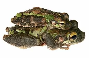
Background imageReproductive Collection: Buckley bonehead frogs mating

Buckley bonehead frogs mating
Buckley bonehead (Osteocephalus buckleyi) frogs mating. This position is known as amplexus. The male frog (top) grasps the female (bottom) with his front legs while she lays her eggs

Background imageReproductive Collection: Woman holding split puffball fungus

Woman holding split puffball fungus
MODEL RELEASED. Woman holding a split puffball fungus. The outer wall of this fruiting body has split open to allow the dispersal of the reproductive spores from the sac inside

Background imageReproductive Collection: Fern leaf, SEM

Fern leaf, SEM
Fern leaf. Coloured scanning electron micrograph (SEM) of the underside of a fern leaf, showing sori. A sorus is a group of sporangia (brown)

Background imageReproductive Collection: Male peacock displaying

Male peacock displaying
Male peacock. Computer enhanced image of a male peacock (Pavo sp.) displaying its tail feathers. The males extravagant feathers are displayed as part of its courtship ritual

Background imageReproductive Collection: Cuckoo Pint (Arum maculatum)

Cuckoo Pint (Arum maculatum). Cross section through the inflorescence

Background imageReproductive Collection: Coot sitting on a nest

Coot sitting on a nest
Female coot (Fulica atra) sitting on a nest in a river

Background imageReproductive Collection: Fern frond, light micrograph

Fern frond, light micrograph. Transverse section through the frond of a fern (Cyrtomium falcatum), showing the sporangia. At left is a central vascular bundle (meristele)

Background imageReproductive Collection: Gorse petal surface, SEM

Gorse petal surface, SEM
Gorse (Ulex europaeus) petal surface, coloured scanning electron micrograph (SEM). This surface is smooth, compared to the surfaces of leafs and sepals

Background imageReproductive Collection: Clubmoss cone, light micrograph

Clubmoss cone, light micrograph. Longitudinal section through the cone (strobilus) of the clubmoss Lycopodium clavatum. The strobilus consists of a central axis (purple)

Background imageReproductive Collection: Fern plant reproduction, light micrograph

Fern plant reproduction, light micrograph. At bottom, the double kidney-shaped gametophyte (prothallus) plant has anchored itself with root-like rhizoids (central black area)

Background imageReproductive Collection: Horsetail cone, light micrograph

Horsetail cone, light micrograph. Longitudinal section through a young cone (strobilus) of the rough horsetail (Equisetum hyemale)

Background imageReproductive Collection: Dandelion pappus, SEM

Dandelion pappus, SEM
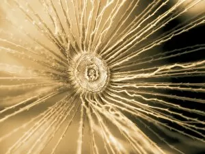
Dandelion pappus (Taraxacum officinale), coloured scanning electron micrograph (SEM). This is the top of one of the hundreds of seeds attached to a dandelion seedhead

Background imageReproductive Collection: Daisy pollen grains, SEM

Daisy pollen grains, SEM
Daisy (Bellis perennis) pollen grains, coloured scanning electron micrograph (SEM). Pollen grains are reproductive structures produced by the male parts of flowering plants

Background imageReproductive Collection: Liverwort spore capsule, light micrograph

Liverwort spore capsule, light micrograph. Longitudinal section through the thallus and sporangium of a liverwort (Jungammania sp.) The sporangium (spore capsule)

Background imageReproductive Collection: Moss reproductive parts, light micrograph

Moss reproductive parts, light micrograph. Longitudinal section through the antheridial cup (male reproductive parts) of a cord moss (Polytrichum commure)

Background imageReproductive Collection: Liverwort spore elaters, light micrograph

Liverwort spore elaters, light micrograph. Transverse section through the sporangium of a liverwort (Pellia epiphylla). This is the basal portion of the sporangium

Background imageReproductive Collection: Bluebell pollen grains, SEM

Bluebell pollen grains, SEM
Bluebell (Hyacinthoides non-scripta) pollen grains, coloured scanning electron micrograph (SEM). Pollen grains are reproductive structures produced by the male parts of flowering plants

Background imageReproductive Collection: Moss spore capsule, light micrograph

Moss spore capsule, light micrograph
Moss spore capsule, polarised light micrograph. Longitudinal section through a spore capsule from a fire moss (Funaria hygrometrica)

Background imageReproductive Collection: Mushroom gill cap, light micrograph

Mushroom gill cap, light micrograph. Transverse section through the cap of a common mushroom (Psalliota sp.) showing the gill lamellae (white)

Background imageReproductive Collection: Plantain pollen grain, SEM

Plantain pollen grain, SEM
Plantain (Plantago lanceolata) pollen grain, coloured scanning electron micrograph (SEM). Pollen grains are reproductive structures produced by the male parts of flowering plants

Background imageReproductive Collection: Liverwort spores, light micrograph

Liverwort spores, light micrograph. Transverse section through the sporangium of a liverwort (Pellia epiphylla). Part of the sporangiums outer wall is at left

Background imageReproductive Collection: Rust fungus infection, light micrograph

Rust fungus infection, light micrograph. Transverse section through a barberry (Berberis sp.) leaf infected by a parasitic rust (Puccinia sp.)

Background imageReproductive Collection: Buttercup pollen with leaf trichome, SEM

Buttercup pollen with leaf trichome, SEM
Buttercup (Ranunculus repens) pollen (orange) with a trichome (leaf hair, green), coloured scanning electron micrograph (SEM)

Background imageReproductive Collection: Glacier lily (Erythronium grandiflorum)

Glacier lily (Erythronium grandiflorum) flowering in the snow. Photographed on Mount Rainier, Cascade Mountains, Washington, USA

Background imageReproductive Collection: Fern spore capsule, light micrograph

Fern spore capsule, light micrograph
Fern spore capsule, polarised light micrograph. Transverse section through the spororocarp (spore capsule) of the water fern pillwort (Pilularia globulifera)

Background imageReproductive Collection: Buttercup pollen grains, SEM

Buttercup pollen grains, SEM
Buttercup (Ranunculus repens) pollen grains, coloured scanning electron micrograph (SEM). Pollen grains are reproductive structures produced by the male parts of flowering plants

Background imageReproductive Collection: Mould fruiting bodies, light micrograph

Mould fruiting bodies, light micrograph. Black mould (Rhizopus nigricans) fruiting bodies (top and upper right) liberating spores

Background imageReproductive Collection: Algal colony, light micrograph

Algal colony, light micrograph. Colony of the green freshwater alga Coleochaete sentata. The whole plant is a disc composed of filaments packed close together, branching out from the centre

Background imageReproductive Collection: Gorse pollen grains, SEM

Gorse pollen grains, SEM
Gorse (Ulex europaeus) pollen grains (yellow), coloured scanning electron micrograph (SEM). Pollen grains are reproductive structures produced by the male parts of flowering plants


All Professionally Made to Order for Quick Shipping

"Exploring the Intricate World of Reproduction: From Dinosaurs to Flowers and Beyond" In the ancient world, even mighty Tyrannosaurus rex dinosaurs had their own unique ways of reproducing. Discovering fossilized evidence of their mating rituals offers a fascinating glimpse into the reproductive strategies employed by these prehistoric giants. Zooming in closer to modern times, microscopic wonders like lavender pollen grains under scanning electron microscopy (SEM) reveal intricate structures that play a crucial role in plant reproduction. Similarly, SEM images of geranium anthers and dahlia flower pollen showcase nature's attention to detail when it comes to ensuring successful pollination. Nature never ceases to amaze with its diverse range mechanisms. Take the cloudberry, for example - this Arctic fruit relies on cross-pollination facilitated by insects or wind for its continued existence. Moving beyond plants, we find ourselves exploring the realm of humans. The miracle of life unfolds within a pregnant woman's body as she nurtures new beginnings and prepares for motherhood. But reproduction is not limited to just larger organisms; even tiny creatures like honeybees have their own unique methods. SEM images capturing honeybee legs provide insight into how these industrious insects transport precious pollen from one flower to another, ensuring fertilization occurs. Venturing further into nature's hidden corners reveals captivating details about reproduction in unexpected places. Mushroom gills under SEM expose delicate structures designed for spore dispersal – a vital step in mushroom reproduction. Delving deeper still, tea flower stamens captured through SEM offer an up-close look at yet another facet of botanical fertility – showcasing intricate arrangements meant to attract pollinators and ensure successful seed production. Flowers continue enchanting us with their beauty as well as their reproductive prowess. Delphinium flowers boast vibrant colors while employing various strategies such as self-pollination or attracting specific pollinators like bees or butterflies.