
Reproductive Collection
"Exploring the Intricate World of Reproduction: From Dinosaurs to Flowers and Beyond" In the ancient world


All Professionally Made to Order for Quick Shipping
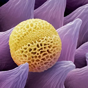
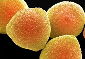
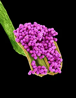
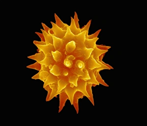
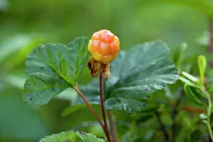
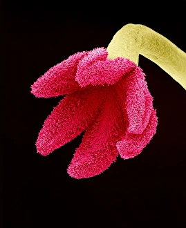
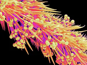
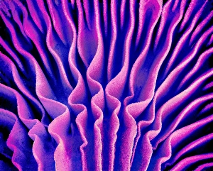
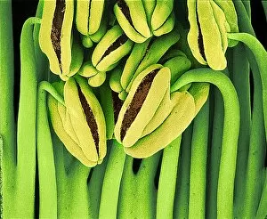
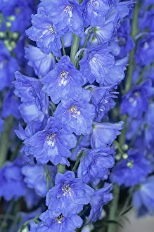
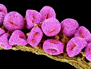
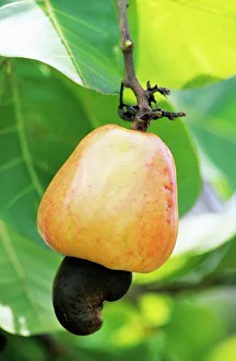
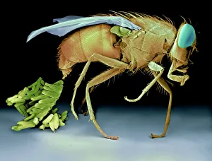
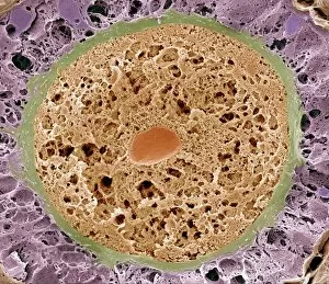
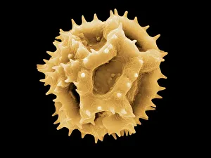
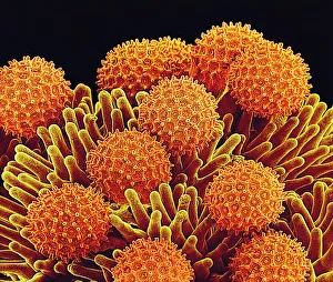
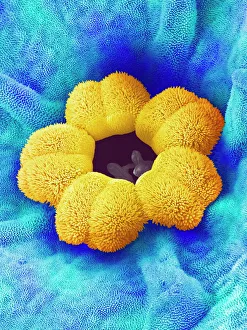
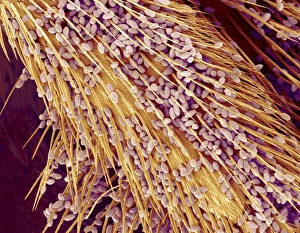
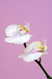
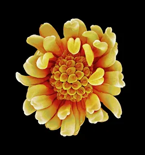
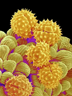
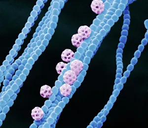
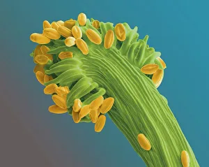
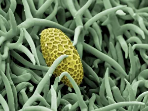
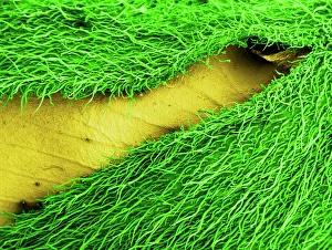
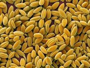
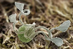
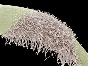
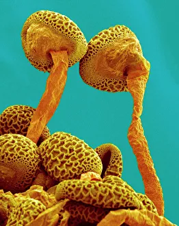
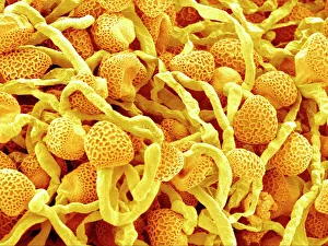
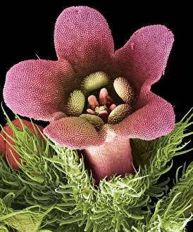
"Exploring the Intricate World of Reproduction: From Dinosaurs to Flowers and Beyond" In the ancient world, even mighty Tyrannosaurus rex dinosaurs had their own unique ways of reproducing. Discovering fossilized evidence of their mating rituals offers a fascinating glimpse into the reproductive strategies employed by these prehistoric giants. Zooming in closer to modern times, microscopic wonders like lavender pollen grains under scanning electron microscopy (SEM) reveal intricate structures that play a crucial role in plant reproduction. Similarly, SEM images of geranium anthers and dahlia flower pollen showcase nature's attention to detail when it comes to ensuring successful pollination. Nature never ceases to amaze with its diverse range mechanisms. Take the cloudberry, for example - this Arctic fruit relies on cross-pollination facilitated by insects or wind for its continued existence. Moving beyond plants, we find ourselves exploring the realm of humans. The miracle of life unfolds within a pregnant woman's body as she nurtures new beginnings and prepares for motherhood. But reproduction is not limited to just larger organisms; even tiny creatures like honeybees have their own unique methods. SEM images capturing honeybee legs provide insight into how these industrious insects transport precious pollen from one flower to another, ensuring fertilization occurs. Venturing further into nature's hidden corners reveals captivating details about reproduction in unexpected places. Mushroom gills under SEM expose delicate structures designed for spore dispersal – a vital step in mushroom reproduction. Delving deeper still, tea flower stamens captured through SEM offer an up-close look at yet another facet of botanical fertility – showcasing intricate arrangements meant to attract pollinators and ensure successful seed production. Flowers continue enchanting us with their beauty as well as their reproductive prowess. Delphinium flowers boast vibrant colors while employing various strategies such as self-pollination or attracting specific pollinators like bees or butterflies.