Fraud Blocker Skip to main content

Radiological Collection (page 2)

"Unlocking the Secrets: Radiological Insights into Our Bodies" Revealing a Healthy Knee

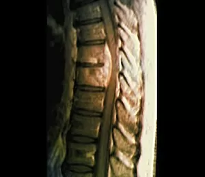
Background imageRadiological Collection: Spondylodiscitis, MRI scan

Spondylodiscitis, MRI scan
Spondylodiscitis. Coloured magnetic resonance imaging (MRI) scan of a sagittal section through the spine of a 69-year-old patient with spondylodiscitis at the level of the T7-T8 vertebra
Background imageRadiological Collection: Abnormal bone growth, MRI scan

Abnormal bone growth, MRI scan
Abnormal bone growth. Magnetic resonance imaging (MRI) scan of a section through the cervical spine (neck) of a 28-year-old patient, showing abnormal growth (dysplasia) of the C6 vertebra

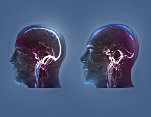
Background imageRadiological Collection: Thrombophlebitis in the brain, MRI scans

Thrombophlebitis in the brain, MRI scans
Thrombophlebitis in the brain. Coloured magnetic resonance imaging (MRI) angiograms comparing the blood vessels (white) of the brain and neck in a healthy 43-year-old (left)

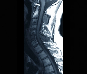
Background imageRadiological Collection: Spinal cancer, MRI scan

Spinal cancer, MRI scan
Spinal cancer. Coloured magnetic resonance imaging (MRI) scan of a sagittal section through the cervical (neck) region of the spine of a 22-year-old patient

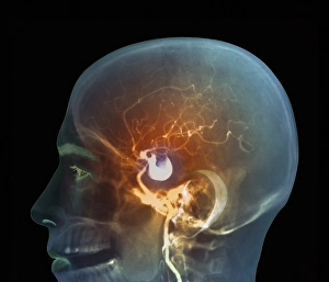
Background imageRadiological Collection: Brain aneurysm, DSA scan

Brain aneurysm, DSA scan
Brain aneurysm. Coloured digital subtraction angiogram (DSA) of the head of a 38-year-old, showing a large aneurysm (bright, centre) of the right internal carotid artery

Background imageRadiological Collection: Thrombophlebitis in the brain, 3D CT scan

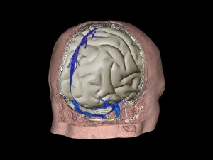
Thrombophlebitis in the brain, 3D CT scan
Thrombophlebitis in the brain. Coloured 3D computed tomography (CT) angiogram of the blood vessels (blue) in the brain of a 48-year-old with thrombophlebitis of the superior sagittal sinus

Background imageRadiological Collection: Gorhams disease, 3D CT scan

Gorhams disease, 3D CT scan
Gorhams disease. Coloured 3D computed tomography (CT) scan of the rear of the skull of a 47-year-old patient, showing damage (centre) caused by osteolysis (bone resorption and replacement)

Background imageRadiological Collection: Forestiers disease, MRI scan

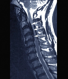
Forestiers disease, MRI scan
Forestiers disease. Magnetic resonance imaging (MRI) scan of a section through the cervical spine (neck) of a 42-year-old Asian patient suffering from a stiff neck and neck pain

Background imageRadiological Collection: Spina bifida, MRI scan

Spina bifida, MRI scan
Fibrolipoma of the spine. Magnetic resonance imaging (MRI) scan of a sagittal section through the spine of a 24-month-old patient with spina bifida

Background imageRadiological Collection: Coronary stenosis after treatment, X-ray

Coronary stenosis after treatment, X-ray
Coronary stenosis after treatment. Angiogram of the left coronary artery in the heart of a 60-year-old patient after balloon catheterisation

Background imageRadiological Collection: Ventricular shunt, X-ray

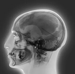
Ventricular shunt, X-ray
Ventricular shunt. X-ray of the head of a 52-year-old patient with hydrocephalus, showing the position of a ventricular shunt (dark lines, right) and its external valve (round, centre right)

Background imageRadiological Collection: Thickened skull, MRI scan

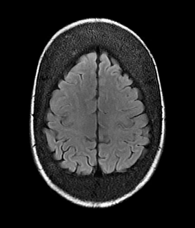
Thickened skull, MRI scan
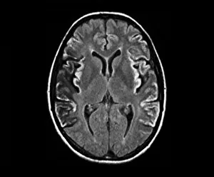
Thickened skull. Magnetic resonance imaging (MRI) scan of an axial section through the head of a 22-year-old patient, showing thickening of the trabecular (spongy) bone of the skull

Background imageRadiological Collection: Secondary spinal cancer, MRI scan

Secondary spinal cancer, MRI scan
Secondary spinal cancer. Magnetic resonance imaging (MRI) scan of a section through the spine of a 48-year-old patient, showing malignant (cancerous) tumours

Background imageRadiological Collection: Gorhams disease, CT scans

Gorhams disease, CT scans
Gorhams disease. Coloured 3D (left) and 2D (right) computed tomography (CT) scans of the skull of a 47-year-old patient, showing damage (centre) caused by osteolysis (bone resorption and replacement)

Background imageRadiological Collection: Stroke, CT scans

Stroke, CT scans
Stroke. Coloured computed tomography (CT) scans of sections through the brain of a patient taken 24 hours, 10 days, 25 days, and 90 days after a haemorrhagic stroke

Background imageRadiological Collection: Coronary stenosis treatment, X-rays

Coronary stenosis treatment, X-rays
Coronary stenosis treatment. Coloured angiograms showing the left coronary artery in the heart of a 60-year-old patient before (top left)

Background imageRadiological Collection: Fibrolipoma of the spine, MRI scan

Fibrolipoma of the spine, MRI scan
Fibrolipoma of the spine. Coloured magnetic resonance imaging (MRI) scan of a sagittal section through the thoraco-lumbar spine of a 19-month-old patient

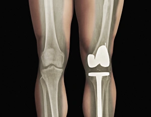
Background imageRadiological Collection: Total knee replacement, X-ray

Total knee replacement, X-ray
Total knee replacement. X-ray of the legs of a 49-year-old patient who has had a total knee replacement (white) of the left knee (right)

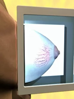
Background imageRadiological Collection: Mammogram, conceptual image

Mammogram, conceptual image. Computer artwork of a womans right breast in an X-ray machine during a mammogram (breast X-ray)

Background imageRadiological Collection: Diagnostics imaging techniques, concept C016 / 7511

Diagnostics imaging techniques, concept C016 / 7511
Diagnostics imaging techniques, conceptual computer artwork

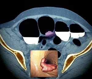
Background imageRadiological Collection: Colon cancer, barium contrast CT scan

Colon cancer, barium contrast CT scan
Colon cancer. Coloured barium contrast computed tomography (CT) scan, and 3D CT colonoscopy image (inset), of an axial section through the sigmoid colon (large intestine) of a 54-year-old patient

Background imageRadiological Collection: Colon cancer, 3D colonoscopy image

Colon cancer, 3D colonoscopy image
Colon cancer. 3D colonoscopy image showing the location of a cancerous growth (centre right) in the colon of a 59-year-old patient. Here, the growth has caused a narrowing (stenosis) of the colon

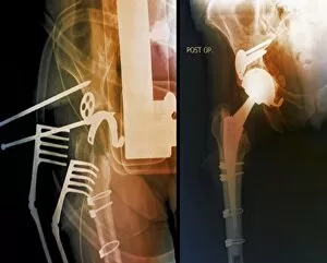
Background imageRadiological Collection: Total hip replacement, X-rays

Total hip replacement, X-rays
Total hip replacement. Coloured X-rays taken during (left) and after (right) surgery to replace the right hip joint of a 65-year-old patient using a prosthetic implant

Background imageRadiological Collection: Hearing aid, X-ray

Hearing aid, X-ray
Hearing aid. Coloured X-ray of the head of a 70-year-old patient, showing a hearing aid (white, centre left) located behind their left ear

Background imageRadiological Collection: Breast lumps, MRI

Breast lumps, MRI
Breast lumps. Coloured magnetic resonance imaging (MRI) scan of the chest of a 20-year-old patient, showing multiple fibroadenomas (light patches) in both breasts

Background imageRadiological Collection: Narrowed aorta, 3D CT scans

Narrowed aorta, 3D CT scans
Narrowed aorta. Coloured 3D computed tomography (CT) scans of the heart (centre of each scan) and major blood vessels of a 50-year-old patient

Background imageRadiological Collection: Targeted breast surgery, mammogram

Targeted breast surgery, mammogram
Targeted breast surgery. Coloured mammograms of the right breast of a 47-year-old female patient being used to locate the exact site of micro-lesions, prior to surgery

Background imageRadiological Collection: Colon polyp, 3D colonoscopy image

Colon polyp, 3D colonoscopy image
Colon polyp. Coloured 3D computed tomography (CT) colonoscopy image of a pedunculated (stalked) polyp in the sigmoid colon (large intestine) of a 50-year-old patient

Background imageRadiological Collection: Failed hip replacement, CT scan

Failed hip replacement, CT scan
Failed hip replacement. Coloured computed tomography (CT) scan of a total hip replacement (solid white) in a 70-year-old patient

Background imageRadiological Collection: Calcified heart valve, CT scan

Calcified heart valve, CT scan
Calcified heart valve. Coloured computed tomography (CT) scan of a section through the heart of a 67-year-old patient showing calcification of the aortic valve (round)

Background imageRadiological Collection: Brain tumour, MRI scans

Brain tumour, MRI scans
Brain tumour. Coloured magnetic resonance imaging (MRI) scans of sections through the head of a 23-year-old patient with a lesion (dark patches)

Background imageRadiological Collection: Stroke, MRI scan

Stroke, MRI scan
Stroke. Coloured magnetic resonance imaging (MRI, left) and computed tomography (CT, right) scans of axial sections through the brain of a 58-year-old patient

Background imageRadiological Collection: Berry aneurysm, MRI scan

Berry aneurysm, MRI scan
Stroke. Coloured computed tomography (CT) scan of an axial section through the brain of a 58-year-old patient, showing the damage (dark) caused 24 hours after a cerebral infarction

Background imageRadiological Collection: Creutzfeldt-Jakob disease, MRI scan

Creutzfeldt-Jakob disease, MRI scan
Creutzfeldt-Jakob disease. Coloured magnetic resonance imaging (MRI) scan of an axial section through the brain of a 35-year-old patient

Background imageRadiological Collection: Stroke, CT scan

Stroke, CT scan
Stroke. Coloured computed tomography (CT) scan of an axial section through the brain of a 58-year-old patient, showing the damage (purple) caused 24 hours after a cerebral infarction

Background imageRadiological Collection: Double mastectomy breast implants, MRI

Double mastectomy breast implants, MRI
Double mastectomy breast implants. Post-operative coloured magnetic resonance imaging (MRI) scan of a section through the chest of a 61-year-old patient after a double mastectomy (breast removal)

Background imageRadiological Collection: Kidney tumour, CT scan

Kidney tumour, CT scan
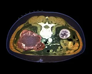
Kidney tumour. Coloured computed tomography (CT) scan of a section through the abdomen of a 62-year-old patient, showing necrosis (death) of tissue in the right kidney

Background imageRadiological Collection: Hip fracture reduction, 3D CT scan

Hip fracture reduction, 3D CT scan
Hip fracture reduction. Coloured 3D computed tomography (CT) scan of the pelvis of an 81-year-old patient with a fracture to the neck of the right femur (thigh bone)

Background imageRadiological Collection: Secondary lung cancer, X-ray

Secondary lung cancer, X-ray
Secondary lung cancers. Coloured X-ray of malignant (cancerous) tumours (red patches) in the lungs of a 60-year-old female patient

Background imageRadiological Collection: Calcified heart valve, CT scans

Calcified heart valve, CT scans
Calcified heart valve. Coloured computed tomography (CT) scans of a section through the heart of a 67-year-old patient showing calcification of the aortic valve (round)

Background imageRadiological Collection: Colon cancer, CT scans

Colon cancer, CT scans
Colon cancer. 2D computed tomography (CT) rendering (left) and 3D colonoscopy image (right) showing the location of a cancerous growth in the colon of a 59-year-old patient


All Professionally Made to Order for Quick Shipping

"Unlocking the Secrets: Radiological Insights into Our Bodies" Revealing a Healthy Knee: A closer look through X-ray imaging showcases the strength and resilience of a healthy knee joint. Unmasking Bacterial Meningitis: An MRI scan provides vital radiological evidence in diagnosing bacterial meningitis, aiding doctors in timely treatment. On-the-Go Imaging Solutions: The advent of mobile x-ray units brings convenience and efficiency to healthcare, ensuring prompt diagnosis and care wherever needed. Restoring Mobility with Total Knee Replacement: X-rays play a crucial role in assessing successful outcomes after total knee replacement surgeries, offering insights into improved mobility for patients. Peering into a Healthy Ankle Joint: Through an x-ray lens, witness the intricate structure of a healthy ankle joint that supports our daily activities without hindrance. Detecting Scoliosis through X-ray Vision: X-rays help identify scoliosis early on, enabling effective treatment plans to alleviate discomfort and maintain spinal health. Mapping Normalcy with Leg X-rays: Explore the fascinating skeletal framework of normal legs captured by x-ray technology – an invaluable tool for orthopedic assessments. Visualizing Ischaemia's Impact with Digital Angiograms: Digital angiograms provide detailed imagery to evaluate blood flow disruptions caused by ischaemia, guiding targeted interventions for better patient outcomes. Decoding Arthrosis of the Hand via X-Ray F006/4616: Delve into the intricacies of arthrosis affecting hand joints using specialized x-rays (F006/4616), aiding accurate diagnoses and personalized treatments. Illuminating Brain Aneurysms through 3D Scans: Cutting-edge 3D scanning techniques shed light on brain aneurysms' complex anatomy, assisting neurosurgeons in planning precise interventions for optimal patient care. Spotting Tuberculosis of the Spine with MRI Scans.