Fraud Blocker Skip to main content

Radiograph Collection (page 6)

"Unveiling the Hidden World: Exploring Radiographs Beyond the Ordinary" Step into a captivating realm where radiographs reveal secrets hidden beneath our very eyes

Background imageRadiograph Collection: Gorilla skull

Gorilla skull (Gorilla). Side view X-ray showing the cranium, eye socket, nasal area and jawbone

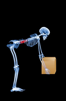
Background imageRadiograph Collection: Skeleton lifting a box incorrectly

Skeleton lifting a box incorrectly. Side view X- ray. This image shows the incorrect method of lifting heavy objects using the lower back muscles with the spine bent
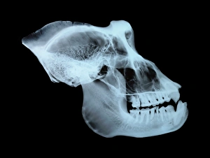
Background imageRadiograph Collection: Monkey skull, X-ray

Monkey skull, X-ray
Monkey skull. X-ray image of the skull of a monkey (species unknown). Elongated front teeth (canines) are found in many monkey species, often in males only

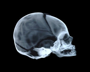
Background imageRadiograph Collection: Babys skull

Babys skull. Side view X-ray showing the cranium, eye socket, nasal area and mouth. For an X-ray of an adult human skull see image P120/222 and for an X-ray of a childs skull see image P120/224

Background imageRadiograph Collection: Normal chest, coloured x-ray C013 / 5283

Normal chest, coloured x-ray C013 / 5283
False-colour normal chest x-ray, showing a womans healthy lungs, elongated heart and the aortic arch. The lungs (yellow-orange areas to the left and right of the central spinal column)

Background imageRadiograph Collection: Normal chest coloured x-ray C013 / 5280

Normal chest coloured x-ray C013 / 5280
False-colour normal chest x-ray, side view, showing a womans healthy lungs (yellow-orange areas to the right of the spinal column)

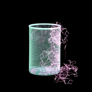
Background imageRadiograph Collection: Earthworms in a can, X-ray

Earthworms in a can, X-ray
Earthworms (Lumbricus sp.) spilling out of a tin can, coloured X-ray

Background imageRadiograph Collection: Angel wing shell, X-ray

Angel wing shell, X-ray
Angel wing shell (Cyrtopleura costata), coloured X-ray. The angel wing is a bivalve marine mollusc. It burrows into coastal sands and feeds on algae through a siphon

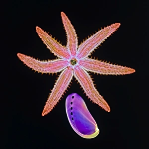
Background imageRadiograph Collection: Starfish and abalone, X-ray

Starfish and abalone, X-ray
Starfish and abalone. Coloured X-ray of a starfish (class Asteroidea) and an abalone (Haliotis sp.). A starfish typically has 5 arms radiating outward from a central disc

Background imageRadiograph Collection: Queen scallop shell, X-ray

Queen scallop shell, X-ray
Queen scallop shell (Chlamys delicatula), coloured X-ray. The queen scallop is a coldwater, bivalve marine mollusc of sub-Antarctic waters

Background imageRadiograph Collection: Swallow, X-ray

Swallow, X-ray
Swallow. Coloured X-ray of a swallow (family Hirundidae). The swallow spends much of its time on the wing, and it is well adapted for energy- efficient flight; its hollow bones

Background imageRadiograph Collection: Gorilla skull, X-ray

Gorilla skull, X-ray
Gorilla skull. Coloured X-ray of a front view of the skull of a male gorilla (Gorilla gorilla). The gorilla is the largest living primate

Background imageRadiograph Collection: Seahorses, X-ray

Seahorses, X-ray
Seahorses, historical X-ray. Some of the seahorses (Hippocampus sp.) are long dead, and have been dried. These appear hollow

Background imageRadiograph Collection: Seahorse, X-ray

Seahorse, X-ray
Seahorse. Coloured X-ray of the external bony skeleton of a seahorse (Hippocampus sp.). The gill cavity is seen behind the eye

Background imageRadiograph Collection: Diagnostic imaging

Diagnostic imaging. Conceptual computer artwork of a human figure with an exposed X-ray plate over his arm and ribcage. This may represent diagnostic medical imaging

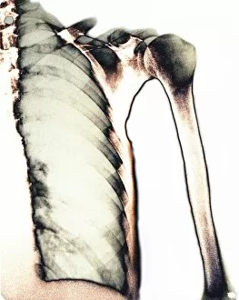
Background imageRadiograph Collection: Healthy lung, X-ray

Healthy lung, X-ray
Healthy lung. Coloured X-ray of a healthy human lung. The spine runs vertically down far left. The ribs curve across the chest and protect the lungs. The upper arm bone (humerus) is at right

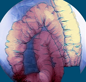
Background imageRadiograph Collection: Large intestine, X-ray

Large intestine, X-ray
Large intestine. Coloured barium X-ray of a section of a healthy large intestine (pink/yellow), the final section of the digestive tract

Background imageRadiograph Collection: Lung bronchogram, coloured X-ray

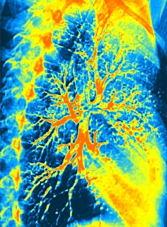
Lung bronchogram, coloured X-ray
Healthy lung. Coloured bronchogram x-ray of a side (lateral) view of a healthy human lung. The vertebrae of the spinal column are seen at left

Background imageRadiograph Collection: Lungs and heart, X-ray

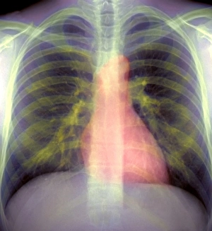
Lungs and heart, X-ray
Lungs and heart, coloured chest X-ray. The lungs and heart are enclosed and protected by the ribs (horizontal bars) which meet at the breastbone, or sternum (down centre)

Background imageRadiograph Collection: Hip bones, X-ray

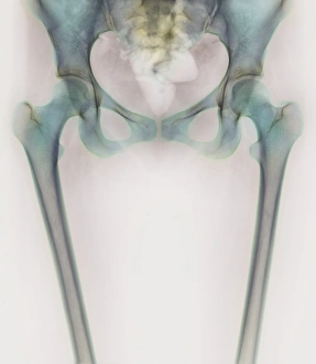
Hip bones, X-ray
Hip joints. Coloured X-ray of a womans groin and upper legs, revealing the bones that form her hip joints. The left hip (identical to the right one) is at upper left in this rear view

Background imageRadiograph Collection: Skeleton stretching

Skeleton stretching. Coloured side view X-ray of a human skeleton performing stretching exercises on an exercise mat

Background imageRadiograph Collection: Coccyx and lower back, X-ray

Coccyx and lower back, X-ray
Coccyx and lower back. Coloured X-ray of a side view of the coccyx and lower back of a healthy adult man. The front of the body is at right

Background imageRadiograph Collection: Skeleton lifting a box correctly

Skeleton lifting a box correctly. Side view X- ray. This image shows the correct method of lifting heavy objects using the leg muscles in a crouched position with the spine straight

Background imageRadiograph Collection: Neck and skull, X-ray

Neck and skull, X-ray
Neck and skull. Coloured X-ray of a normal human neck (lower centre) and part of the skull (upper centre). The neck joins the head to the body

Background imageRadiograph Collection: Knee joint, X-ray

Knee joint, X-ray
Knee joint. Coloured X-ray of the healthy knee joint of a 15-year-old girl. The knee joint is composed of three bones; the femur (thigh bone, upper frame), the tibia (shin bone)

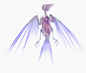
Background imageRadiograph Collection: Flying skeleton

Flying skeleton, computer enhanced X-ray

Background imageRadiograph Collection: Calculator, simulated X-ray

Calculator, simulated X-ray
Calculator. Simulated X-ray of a calculator

Background imageRadiograph Collection: CD drive, simulated X-ray

CD drive, simulated X-ray
CD drive. Simulated X-ray of a compact disc (CD) drive from a computer. A disc is in the sliding tray at bottom. This will retract into the machine

Background imageRadiograph Collection: Computer keyboard, simulated X-ray

Computer keyboard, simulated X-ray
Computer keyboard. Simulated X-ray of a keyboard for a personal computer (PC). The keyboard connects to a computer (not seen) by the lead at upper right

Background imageRadiograph Collection: Computer hard disk, simulated X-ray

Computer hard disk, simulated X-ray
Hard disk. Simulated X-ray of a hard disk memory mechanism from a computer. This consists of a read/write head (pointed, black) on a rigid hard disk (upper centre)

Background imageRadiograph Collection: Purse, simulated X-ray

Purse, simulated X-ray
Purse. Simulated X-ray of a purse containing British pound sterling currency. The ten pound note bears an image of Queen Elizabeth II

Background imageRadiograph Collection: CD drive, coloured X-ray

CD drive, coloured X-ray
CD drive. Coloured X-ray of a compact disc (CD) drive from a computer. A disc is in the sliding tray at bottom. This will retract into the machine

Background imageRadiograph Collection: Computer, simulated X-ray

Computer, simulated X-ray
Computer. Simulated X-ray of a vertical processing unit of a computer. The front of the unit (right) contains removable disc drives for data storage

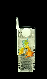
Background imageRadiograph Collection: Mobile phone X-ray

Mobile phone X-ray with a coloured map of the British Isles superimposed. This conceptual image could represent regional variation in patterns of mobile phone usage or network coverage

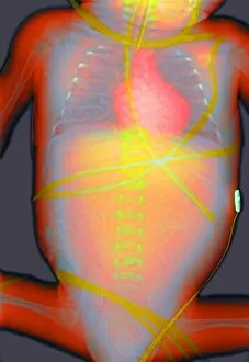
Background imageRadiograph Collection: Neonatal infant, X-ray

Neonatal infant, X-ray
Neonatal infant. Coloured X-ray of a one month old baby. The babys spine (down centre), heart (pink, upper centre) and ribs (upper left and right) are seen. Various life support devices are visible

Background imageRadiograph Collection: Dental X-ray

Dental X-ray
MODEL RELEASED. Dental X-ray. Dentist looking at an X-ray of a patients teeth. This is a panoramic wraparound X-ray that shows all the teeth of both the upper and lower jaw

Background imageRadiograph Collection: Blocked Fallopian tubes, X-ray

Blocked Fallopian tubes, X-ray
Blocked Fallopian tubes. Coloured X-ray of a womans reproductive system, with her uterus (green) and blocked Fallopian tubes (oviducts, yellow)

Background imageRadiograph Collection: Double hip replacement, X-ray

Double hip replacement, X-ray
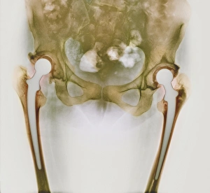
Double hip replacement. Coloured X-ray of a female patients hips with two prosthetic hip implants (white). Each implant is embedded in a hole drilled into its femur (thigh bone)

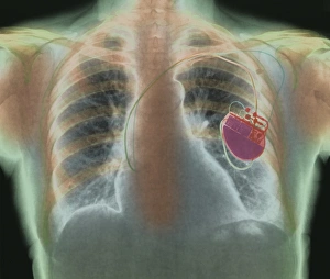
Background imageRadiograph Collection: Heart pacemaker, X-ray

Heart pacemaker, X-ray
Heart pacemaker. Coloured chest X-ray showing a surgically-implanted heart pacemaker (red, centre right). The pacemaker supplies electrical impulses along leads (running to lower centre)

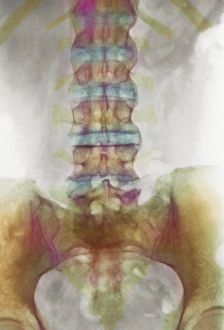
Background imageRadiograph Collection: Spondylitis, X-ray

Spondylitis, X-ray
Spondylitis. Coloured X-ray of the lower spine and pelvis of a patient with spondylitis, an inflammation of the synovial joints of the backbone

Background imageRadiograph Collection: Sarcoidosis of the lungs, X-ray

Sarcoidosis of the lungs, X-ray
Sarcoidosis. Frontal coloured chest X-ray of the lungs of a patient with pulmonary sarcoidosis. Affected lung tissue shows up as red patches

Background imageRadiograph Collection: Thalassaemia, X-ray

Thalassaemia, X-ray
Thalassaemia. Coloured hand X-ray of a patient with thalassaemia, also known as Cooleys anaemia. The bones of the hand show demineralization (lighter patches)

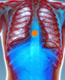
Background imageRadiograph Collection: Swallowed coin, X-ray

Swallowed coin, X-ray
Swallowed coin. Coloured X-ray of a coin (orange) in a childs oesophagus. The heart (blue, centre), ribcage (upper centre) and abdominal region (blue, lower centre) are all seen

Background imageRadiograph Collection: Lung scarring from tuberculosis, X-ray

Lung scarring from tuberculosis, X-ray
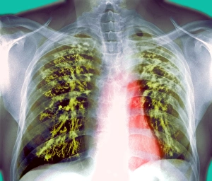
Tuberculosis lung scarring. Coloured chest X-ray showing scarring (green) of the lungs after a case of chronic tuberculosis (TB). The heart (red) is also seen

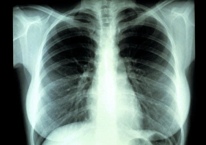
Background imageRadiograph Collection: Lungs, X-ray

Lungs, X-ray

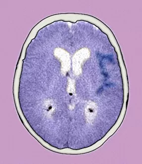
Background imageRadiograph Collection: Brain haemorrhage, CT scan

Brain haemorrhage, CT scan

Background imageRadiograph Collection: Diverticular disease, X-ray

Diverticular disease, X-ray
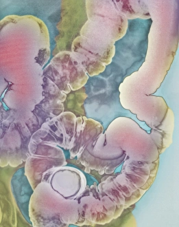
Diverticular disease, coloured X-ray. This illness is also known as diverticulitis or CDD (colonic diverticular disease). This is a double-contrast X-ray

Background imageRadiograph Collection: Paranasal sinuses, X-ray

Paranasal sinuses, X-ray
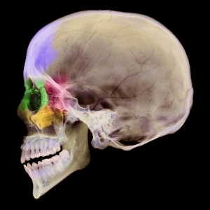
Paranasal sinuses. Coloured X-ray of a sagittal section through a human skull. The skull has been sliced in half down the centre and the regions of the paranasal sinuses coloured


All Professionally Made to Order for Quick Shipping

"Unveiling the Hidden World: Exploring Radiographs Beyond the Ordinary" Step into a captivating realm where radiographs reveal secrets hidden beneath our very eyes. From panoramic dental X-rays capturing intricate details of teeth to seahorse skeletons showcasing nature's delicate beauty, these images offer a glimpse into the mesmerizing world of radiography. Delving deeper, we encounter an ordinary foot transformed by an extraordinary X-ray, exposing its inner structure with remarkable precision. A human skull emerges from darkness, unveiling its intricate contours and reminding us of our mortality. Meanwhile, a horse skull stands as a testament to strength and grace in both life and death. But it doesn't stop there - bunions caught on an X-ray remind us that even seemingly small imperfections can leave their mark. In another twist, irregular heartbeats are captured through this powerful imaging technique, shedding light on conditions that affect millions worldwide. Shifting gears towards artistry within science, we witness an MRI-style X-ray revealing a leg adorned in stiletto shoes – merging fashion with medical technology in stunning detail. Parsley springs to life under the gaze of radiographic vision while tulips and gladiolus showcase their vibrant petals through translucent layers. And finally, behold the Oriental stargazer lily – its ethereal beauty enhanced by colored X-rays that transform it into a work of art beyond imagination. Radiographs have transcended mere diagnostic tools; they have become windows into worlds unseen. As we explore these captivating images together, let us marvel at the wonders revealed by this incredible fusion of science and artistry.