Fraud Blocker Skip to main content

Phytoplankton Collection (page 4)

Phytoplankton, the microscopic wonders of our aquatic world, never cease to amaze us with their intricate beauty and vital role in our ecosystem

Background imagePhytoplankton Collection: Marine Diatoms and Radiolarians

Marine Diatoms and Radiolarians
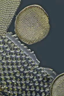
LRMC-17 Scanning Electron Micrograph (SEM): Marine Diatoms and Radiolaria skeletons Magnification x435 (when printed A4, 29.7 cm cm wide)

Background imagePhytoplankton Collection: Marine Diatom

Marine Diatom
LRDS-293 Scanning Electron Micrograph (SEM): Marine Diatom Magnification x2, 570 (when printed 10.5 cm wide) Bacteriastrum sp David Spears (Last Refuge) / ardea.com Last Refuge contact details

Background imagePhytoplankton Collection: Marine Diatom

Marine Diatom
LRDS-291 Scanning Electron Micrograph (SEM): Marine Diatom Magnification x1, 510 (when printed 10.5 cm wide) David Spears (Last Refuge) / ardea.com Last Refuge contact details: prints@ardea.com tel

Background imagePhytoplankton Collection: Sea foam washing up on beach after storm, Hengistbury Head, Dorset, England, april

Sea foam washing up on beach after storm, Hengistbury Head, Dorset, England, april

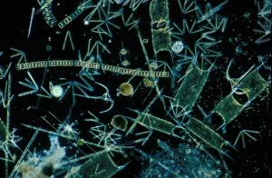
Background imagePhytoplankton Collection: Plankton-Phytopl'ton Phytoplankton x 30 / various diatoms / dino-flagellate etc / English Channel

Plankton-Phytopl'ton Phytoplankton x 30 / various diatoms / dino-flagellate etc / English Channel

Background imagePhytoplankton Collection: Plankton - Phytoplankton / Winter / x30 / coscinodiscus grani /Biddulphia regia

Plankton - Phytoplankton / Winter / x30 / coscinodiscus grani /Biddulphia regia

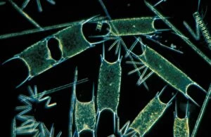
Background imagePhytoplankton Collection: Plankton - x 30 Phytoplankton: Various species of diatom eps. Biddilphia sinensis

Plankton - x 30 Phytoplankton: Various species of diatom eps. Biddilphia sinensis

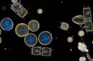
Background imagePhytoplankton Collection: Plankton-Phytopl'ton X20 / various species of diatom & dinoflagellates / English Channel

Plankton-Phytopl'ton X20 / various species of diatom & dinoflagellates / English Channel

Background imagePhytoplankton Collection: Ciliate protozoa, light micrograph

Ciliate protozoa, light micrograph
Ciliate protozoa. Light micrograph of a mixed population of freshwater ciliate protozoa and algae. The algae are the green filaments crossing the field

Background imagePhytoplankton Collection: Calcareous phytoplankton fossil, artwork

Calcareous phytoplankton fossil, artwork
Calcareous phytoplankton fossil. Artwork of a fossil of the skeleton (coccosphere) of a coccolithotrope, a small marine algal organism

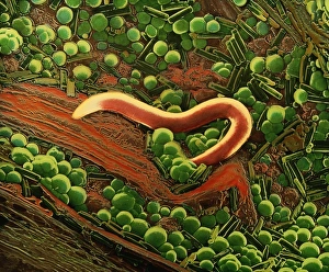
Background imagePhytoplankton Collection: False col SEM of nematode worm on sample of peat

False col SEM of nematode worm on sample of peat
False-colour scanning electron micrograph of a nematode worm on a sample of peat. The micrograph shows the layer of " green slime" which often appears on peat & other organic surfaces

Background imagePhytoplankton Collection: Plankton cell wall, SEM

Plankton cell wall, SEM
Plankton cell wall. Coloured scanning electron micrograph (SEM) of the geometrical plates making up the mineralised cell wall of a planktonic alga (Coronosphaera mediterranea)

Background imagePhytoplankton Collection: Calcareous phytoplankton plates, SEM

Calcareous phytoplankton plates, SEM
Calcareous phytoplankton plates. Coloured scanning electron micrograph (SEM) of parts of the skeleton (coccosphere) of the coccolithophore Emiliania huxleyi, a small marine algal organism

Background imagePhytoplankton Collection: Aquatic crustaceans, light micrograph

Aquatic crustaceans, light micrograph
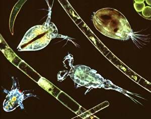
Aquatic crustaceans. Composite image of light micrographs of aquatic planktonic crustaceans with strands of filamentous algae

Background imagePhytoplankton Collection: Rhabdonema marine diatom, SEM

Rhabdonema marine diatom, SEM
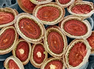
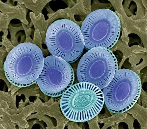
Rhabdonema. Coloured scanning electron micrograph (SEM) of a colony of the marine diatom Rhabdonema sp. Magnification: x500 when printed at 10 centimetres wide

Background imagePhytoplankton Collection: Dried algae

Dried algae on finger produced by Greenfuel Technologies, USA. Algae are photosynthesising microorganisms that convert carbon dioxide (CO2) and sunlight into lipids, carbohydrates and proteins

Background imagePhytoplankton Collection: Algae growing in a photobioreactor

Algae growing in a photobioreactor with a researchers shadow. Algae are photosynthesising micro-organisms that can convert carbon dioxide into commercially valuable lipids

Background imagePhytoplankton Collection: Algae growing on petri dishes

Algae growing on petri dishes, Solazyme Inc. USA. Algae are photosynthesising micro-organisms. They convert carbon dioxide (CO2) into sugars, that are then metabolised into lipids

Background imagePhytoplankton Collection: Algae research

Algae research. Researcher from Solazyme Inc. USA, examining a petri dish from an incubation room containing different algae strains. Algae are photosynthesising micro-organisms

Background imagePhytoplankton Collection: Algae fermentation bioreactor

Algae fermentation bioreactor, Solazyme Inc. USA. Algae are photosynthesising micro- organisms that can convert carbon dioxide into sugars

Background imagePhytoplankton Collection: Researcher with algae fermenter

Researcher with algae fermenter
Researcher with algae fermentation bioreactor, Solazyme Inc. USA. Algae are photosynthesising micro-organisms that can convert carbon dioxide (CO2) into sugars

Background imagePhytoplankton Collection: Atmosphere engineering

Atmosphere engineering. Diagram showing the technology that could be employed in altering the Earths atmospheric conditions to reduce climate change effects

Background imagePhytoplankton Collection: False-col satellite image of worlds oceans

False-col satellite image of worlds oceans
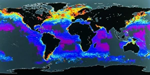
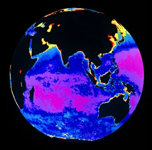
False-colour satellite image of the worlds oceans, showing the distribution of phytoplankton in the surface water. The colours represent varying phytoplankton densities from red (most dense)

Background imagePhytoplankton Collection: False colour image of the Indian Ocean

False colour image of the Indian Ocean
False-colour satellite image of the Indian Ocean and part of the Pacific Ocean, showing the distribution of phytoplankton in the surface water

Background imagePhytoplankton Collection: Diatoms, light micrograph

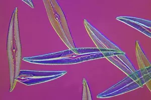
Diatoms, light micrograph. Diatoms are a group of photosynthetic, single-celled algae containing about 10, 000 species. They form an important part of the plankton at the base of the marine

Background imagePhytoplankton Collection: Melosira sp diatoms, light micrograph

Melosira sp diatoms, light micrograph
Diatoms. Light micrograph of Melosira monoliformis freshwater diatoms. Diatoms are a group of photosynthetic, single-celled algae containing about 10, 000 species

Background imagePhytoplankton Collection: Pleurosigma sp diatoms, light micrograph

Pleurosigma sp diatoms, light micrograph
Diatoms. Differential interference contrast micrograph of Pleurosigma angulatum marine diatoms. Diatoms are a group of photosynthetic, single-celled algae containing about 10, 000 species

Background imagePhytoplankton Collection: Fossil diatoms, light micrograph

Fossil diatoms, light micrograph. Diatoms are a group of photosynthetic, single-celled algae containing about 10, 000 species

Background imagePhytoplankton Collection: Pleurosigma sp diatom, light micrograph

Pleurosigma sp diatom, light micrograph
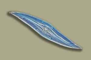
Diatom. Light micrograph of Pleurosigma angulatum marine diatoms. Diatoms are a group of photosynthetic, single-celled algae containing about 10, 000 species


All Professionally Made to Order for Quick Shipping

Phytoplankton, the microscopic wonders of our aquatic world, never cease to amaze us with their intricate beauty and vital role in our ecosystem. From the mesmerizing art of diatom algae captured by Ernst Haeckel to the stunning images of calcareous phytoplankton under scanning electron microscopy (SEM), these tiny organisms hold a wealth of secrets waiting to be explored. Diatoms, one of the most abundant types of phytoplankton, reveal themselves through SEM as delicate structures adorned with intricate patterns. Their cell walls, meticulously observed at high magnification, showcase an astonishing level of detail that rivals any masterpiece created by human hands. These diatoms are not only found in marine environments but also thrive in freshwater habitats like ponds where they contribute to the vibrant tapestry of pond life. The significance extends beyond their aesthetic appeal; they play a crucial role in global carbon cycling and oxygen production. Emiliana huxleyi, a coccolithophore species among them, forms beautiful calcite plates known as coccoliths that can be seen under SEM. These tiny fossils provide valuable insights into Earth's history and evolution. As we delve deeper into understanding these remarkable organisms through advanced imaging techniques such as SEM, we uncover more about their ecological importance and potential applications for various industries including biotechnology and biofuel production. The study and preservation of fossilized diatoms further aid scientists in deciphering past environmental conditions while shedding light on future climate change scenarios. In this captivating journey through microcosms unseen by the naked eye lies an appreciation for nature's intricacy and resilience. Phytoplankton remind us that even within seemingly insignificant beings lie profound contributions to sustaining life on our planet – a reminder worth cherishing as we strive towards protecting our fragile ecosystems for generations to come.