Fraud Blocker Skip to main content

Pelvic Collection (page 2)

"The Pelvic: A Fascinating Intersection of Art, Anatomy, and Health" The pelvic region has captivated artists and scientists alike throughout history

Background imagePelvic Collection: Hip fracture reduction, 3D CT scan

Hip fracture reduction, 3D CT scan
Hip fracture reduction. Coloured 3D computed tomography (CT) scan of the pelvis of an 81-year-old patient with a fracture to the neck of the right femur (thigh bone)

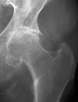
Background imagePelvic Collection: Osteoarthritis of the hip, X-ray C018 / 0593

Osteoarthritis of the hip, X-ray C018 / 0593
Osteoarthritis of the hip. X-ray the left hip of a 74 year old patient showing osteoarthritis. The ball at the top of the femur (thigh bone) fits into the socket of the hip

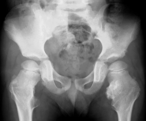
Background imagePelvic Collection: Bone growths, X-ray C018 / 0580

Bone growths, X-ray C018 / 0580
Bone growths. X-ray of the pelvis of a 12 year old patient with bony growths (exostoses) and cartilage cysts (enchondromas) on the femurs (thigh bones) and hip bones

Background imagePelvic Collection: Hip joint bones and anatomy, artwork C014 / 2031

Hip joint bones and anatomy, artwork C014 / 2031
Hip joint bones and anatomy. Artwork of the bones that form the two hip joints (right and left). The hip is a ball-and-socket joint that provides the mobility needed for walking and running

Background imagePelvic Collection: Uterine fibroid, X-ray C014 / 4919

Uterine fibroid, X-ray C014 / 4919
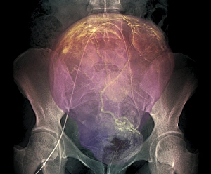
Uterus tumour. Coloured X-ray of the abdomen of a woman with a fibroid (round, centre) in her uterus. A fibroid is a fibrous benign tumour originating from muscular tissue (leimyoma)

Background imagePelvic Collection: Uterine fibroid, X-ray C014 / 4918

Uterine fibroid, X-ray C014 / 4918
Uterus tumour. Coloured X-ray of the abdomen of a woman with a fibroid (orange, round) in her uterus. A fibroid is a fibrous benign tumour originating from muscular tissue (leimyoma)

Background imagePelvic Collection: Endometriosis, X-ray C014 / 4917

Endometriosis, X-ray C014 / 4917
Endometriosis. Coloured X-ray of the abdomen of woman with endometriosis. Endometriosis is the movement of cells from the lining of the uterus to the abdominal cavity

Background imagePelvic Collection: Endometriosis, X-ray C014 / 4916

Endometriosis, X-ray C014 / 4916
Endometriosis. Coloured X-ray of the abdomen of woman with endometriosis. Endometriosis is the movement of cells from the lining of the uterus to the abdominal cavity

Background imagePelvic Collection: Lateral trunk muscles, 1831 artwork

Lateral trunk muscles, 1831 artwork
Lateral trunk muscles. Side view (front of body at right) of the third layer of the lateral muscles of the trunk. The muscles include the serratus, the transverse abdominal

Background imagePelvic Collection: Deep back muscles, 1831 artwork

Deep back muscles, 1831 artwork
Deep back muscles. General disposition of the third layer of muscles in the back, with the base of the skull at top, the ribs at centre, and the pelvis at bottom

Background imagePelvic Collection: Thigh muscle anatomy, 1831 artwork

Thigh muscle anatomy, 1831 artwork
Thigh muscle anatomy. Anterior view of some of the layers of the muscles of the thigh in relation to the femur (thigh bone) and the pelvis bones

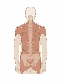
Background imagePelvic Collection: Back muscles, anatomical artwork

Back muscles, anatomical artwork
Back muscles. Anatomical computer artwork showing the muscles of the human back, and shoulders

Background imagePelvic Collection: Hip ligaments, artwork

Hip ligaments, artwork
Hip ligaments. Computer artwork of a human hip, showing the four ligaments (purple) that connect the femur (thigh bone, lower right) to the pelvis to support the hip joint

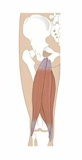
Background imagePelvic Collection: Adductor muscles, artwork

Adductor muscles, artwork
Adductor muscles, computer artwork. Rear view of a human upper leg and right hip (top), showing the structure of the right adductor muscles

Background imagePelvic Collection: Hamstring anatomy, artwork

Hamstring anatomy, artwork
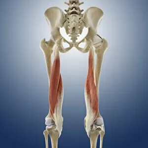
Hamstring anatomy, computer artwork. Rear view of a human upper leg and right hip (top), showing the structure of the right hamstring

Background imagePelvic Collection: Pelvis, artwork C014 / 5105

Pelvis, artwork C014 / 5105
Pelvis. Computer artwork of the pelvis showing the ligaments (white) and nerves (yellow). Ligaments are bands of connective tissue that connect bones

Background imagePelvic Collection: Cardiovascular system, artwork C013 / 5946

Cardiovascular system, artwork C013 / 5946
Cardiovascular system, computer artwork. The heart (centre) is a hollow muscle that pumps blood around the body. Arteries carry oxygenated blood (red) around the body

Background imagePelvic Collection: Hip pain, artwork C013 / 5924

Hip pain, artwork C013 / 5924
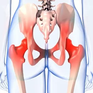
Hip pain, computer artwork

Background imagePelvic Collection: Lower back pain, artwork C013 / 5922

Lower back pain, artwork C013 / 5922
Lower back pain, computer artwork

Background imagePelvic Collection: Hip pain, artwork C013 / 5918

Hip pain, artwork C013 / 5918
Hip pain, computer artwork

Background imagePelvic Collection: Dislocated hip replacement, X-ray C017 / 7804

Dislocated hip replacement, X-ray C017 / 7804
Dislocated hip replacement. X-ray of the pelvis of a patient whos total hip replacement of the rigt hip (left) has become dislocated

Background imagePelvic Collection: Dislocated hip replacement, X-ray C017 / 7803

Dislocated hip replacement, X-ray C017 / 7803
Dislocated hip replacement. X-ray of the pelvis of a patient whos total hip replacement of the rigt hip (left) has become dislocated

Background imagePelvic Collection: Uterine bleeding treatment, DSA scan

Uterine bleeding treatment, DSA scan
Uterine bleeding treatment. Coloured digital subtraction angiogram (DSA) of the pelvic region of a 45-year-old woman with menometrorrhagia, showing embolisation of the uterine arteries

Background imagePelvic Collection: Lower back pain, artwork C013 / 8810

Lower back pain, artwork C013 / 8810
Lower back pain, computer artwork

Background imagePelvic Collection: Hip pain, artwork C013 / 7803

Hip pain, artwork C013 / 7803
Hip pain, computer artwork

Background imagePelvic Collection: Hip pain, artwork C013 / 7802

Hip pain, artwork C013 / 7802
Hip pain, computer artwork

Background imagePelvic Collection: Hip pain, artwork C013 / 7801

Hip pain, artwork C013 / 7801
Hip pain, computer artwork

Background imagePelvic Collection: Thigh muscles, artwork C013 / 4568

Thigh muscles, artwork C013 / 4568
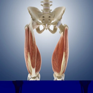
Thigh muscles. Computer artwork of an anterior view of some of the muscles and tendons (red and white) of the thighs and their attachment to the bones of the leg

Background imagePelvic Collection: Thigh muscles, artwork C013 / 4569

Thigh muscles, artwork C013 / 4569
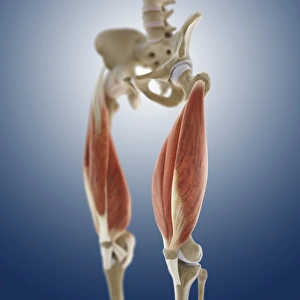
Thigh muscles. Computer artwork of an oblique anterior view of some of the muscles and tendons (red and white) of the thighs and their attachment to the bones of the leg

Background imagePelvic Collection: Hip ligaments, artwork C013 / 4434

Hip ligaments, artwork C013 / 4434
Hip ligaments. Computer artwork of a posterior oblique view of the hip bones and ligaments (white). At upper right the lumbar spine ends in the sacrum and coccyx (tailbone)

Background imagePelvic Collection: Hip ligaments, artwork C013 / 4433

Hip ligaments, artwork C013 / 4433
Hip ligaments. Computer artwork of a posterior view of the hip bones and ligaments (white). At centre the lumbar spine ends in the sacrum and coccyx (tailbone)

Background imagePelvic Collection: Hip ligaments, artwork C013 / 4432

Hip ligaments, artwork C013 / 4432
Hip ligaments. Computer artwork of an anterior view of the hip bones and ligaments (white). At centre the lumbar spine ends in the sacrum and coccyx (tailbone)

Background imagePelvic Collection: Hip anatomy, artwork C013 / 4430

Hip anatomy, artwork C013 / 4430
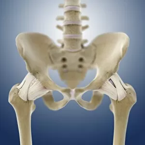
Hip anatomy. Computer artwork of the head of the left femur (centre right) articulating with the left-hand side of the pelvis to form the hip joint
Background imagePelvic Collection: Leg muscles, artwork C013 / 4429

Leg muscles, artwork C013 / 4429
Leg muscles. Computer artwork of the bones and outer muscles of the left leg, seen from the front (anterior view) at an oblique angle looking downwards

Background imagePelvic Collection: Thigh hamstring muscles, artwork C013 / 4428

Thigh hamstring muscles, artwork C013 / 4428
Thigh hamstring muscles. Computer artwork of a posterior view of two of the three hamstring muscles (red) of the thighs. The muscles are the semimembranosus (inner) and semitendinosus (outer)

Background imagePelvic Collection: Iliopsoas muscles, artwork C013 / 4425

Iliopsoas muscles, artwork C013 / 4425
Iliopsoas muscles. Computer artwork of an anterior (frontal) view of the hips, showing the inner hip muscles (red), the ilioposas

Background imagePelvic Collection: Buttock muscles, artwork C013 / 4423

Buttock muscles, artwork C013 / 4423
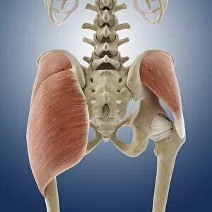
Buttock muscles. Computer artwork of a posterior oblique view of the hips, showing some of the muscles (red) of the buttocks

Background imagePelvic Collection: Buttock muscles, artwork C013 / 4424

Buttock muscles, artwork C013 / 4424
Buttock muscles. Computer artwork of a posterior view of the hips showing some of the muscles (red) of the buttocks. These are shown attached to the femurs (thigh bones) and the pelvis bones

Background imagePelvic Collection: Buttock muscles, artwork C013 / 4422

Buttock muscles, artwork C013 / 4422
Buttock muscles. Computer artwork of a posterior oblique view of the hips showing some of the muscles (red) of the buttocks. These are shown attached to the femurs (thigh bones) and the pelvis bones

Background imagePelvic Collection: Buttock muscles, artwork C013 / 4420

Buttock muscles, artwork C013 / 4420
Buttock muscles. Computer artwork of a posterior view of the hips showing some of the muscles (red) of the buttocks. These are shown attached to the femurs (thigh bones) and the pelvis bones

Background imagePelvic Collection: Buttock muscles, artwork C013 / 4421

Buttock muscles, artwork C013 / 4421
Buttock muscles. Computer artwork of a side view (front of body at left) of the hips showing some of the muscles (red) of the buttocks

Background imagePelvic Collection: Buttock muscles, artwork C013 / 4419

Buttock muscles, artwork C013 / 4419
Buttock muscles. Computer artwork of a posterior oblique view of the hips showing some of the muscles (red) of the buttocks. These are shown attached to the femurs (thigh bones) and the pelvis bones

Background imagePelvic Collection: Buttock muscles, artwork C013 / 4415

Buttock muscles, artwork C013 / 4415
Buttock muscles. Computer artwork of a posterior oblique view of the hips showing some of the muscles (red) of the buttocks. These are shown attached to the femurs (thigh bones) and the pelvis bones

Background imagePelvic Collection: Buttock muscles, artwork C013 / 4416

Buttock muscles, artwork C013 / 4416
Buttock muscles. Computer artwork of a posterior oblique view of the hips showing some of the muscles (red) of the buttocks. These are shown attached to the femurs (thigh bones) and the pelvis bones

Background imagePelvic Collection: Favonigobius sp. goby

Favonigobius sp. goby
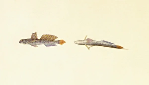
Plate 32A from Zoological drawings by Ferdinand Bauer. Study detail of head, body and tail

Background imagePelvic Collection: Periophthalmus sp. mudskipper

Periophthalmus sp. mudskipper
Plate 32 from Zoological drawings by Ferdinand Bauer. Gobies are common in shallow marine, brackish and estuarine waters, they have a distinct pelvic sucker used to cling to rocks and corals

Background imagePelvic Collection: Hip bones, X-ray

Hip bones, X-ray
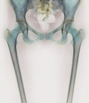
Hip joints. Coloured X-ray of a womans groin and upper legs, revealing the bones that form her hip joints. The left hip (identical to the right one) is at upper left in this rear view

Background imagePelvic Collection: Coccyx and lower back, X-ray

Coccyx and lower back, X-ray
Coccyx and lower back. Coloured X-ray of a side view of the coccyx and lower back of a healthy adult man. The front of the body is at right


All Professionally Made to Order for Quick Shipping

"The Pelvic: A Fascinating Intersection of Art, Anatomy, and Health" The pelvic region has captivated artists and scientists alike throughout history. From ancient historical artwork depicting the cardiovascular system's connection to the pelvis to modern depictions of hip replacement surgeries, this area holds immense significance. One common ailment that affects the pelvic region is osteoarthritis of the hip. X-ray images reveal its impact on the bones, prompting advancements in treatments like hip replacements. These procedures have revolutionized healthcare by restoring mobility and alleviating pain for countless individuals. Another condition associated with the pelvis is a slipped disc. Artists have beautifully portrayed this issue through intricate artworks showcasing slipped intervertebral discs. Such visuals help medical professionals understand and treat this painful condition effectively. Delving deeper into anatomy, X-rays provide detailed views of hip bones from various angles. The complexity of these structures becomes evident as we explore their connections to muscles such as Levator ani - an engraving displaying a side view of the pelvis highlights this relationship. The wonderment surrounding life begins within the womb, where diagrams depict foetuses developing within their mothers' pelvises. This glimpse into human reproduction showcases how essential a healthy pelvic structure is for nurturing new life. Artists have also contributed to our understanding of skeletal anatomy by creating stunning artwork portraying human skeletons in intricate detail. By studying these pieces, we gain insights into how different parts interact within the body's framework. Amidst all these scientific explorations lies an unexpected surprise – a blacktip reef shark. Though not directly related to human anatomy or health concerns involving the pelvic region, it serves as a reminder that nature's wonders extend beyond our own bodies. Returning back to humans, 1831 artwork focuses on another crucial aspect: pelvic-femoral muscles. Understanding their functions aids in comprehending movement patterns and potential issues that may arise when they are compromised. Lastly, artistic representations shed light on the complexity of hip joint bones and their anatomy.