Fraud Blocker Skip to main content

Pathogenic Collection (page 9)

Pathogenic microorganisms are microscopic entities that pose a significant threat to human health

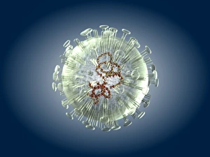
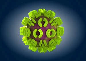
Background imagePathogenic Collection: HIV particle, artwork C018 / 2899

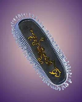
HIV particle, artwork C018 / 2899
HIV particle, computer artwork. HIV (human immunodeficiency virus) causes AIDS (acquired immune deficiency syndrome). The virus consists of an RNA (ribonucleic acid) genome

Background imagePathogenic Collection: Ash dieback disease fungus, SEM C017 / 8028

Ash dieback disease fungus, SEM C017 / 8028
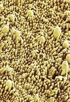
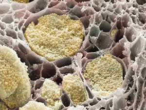
Ash dieback disease fungus. Coloured scanning electron micrograph (SEM) of the surface of a fruiting body of the fungus Chalara fraxinea

Background imagePathogenic Collection: Ash dieback disease fungus, SEM C017 / 8026

Ash dieback disease fungus, SEM C017 / 8026
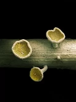
Ash dieback disease fungus. Coloured scanning electron micrograph (SEM) of a fruiting body of the fungus Chalara fraxinea, also known as Hymenoscyphus pseudoalbidus in its fruiting stage

Background imagePathogenic Collection: Ash dieback disease fungus, SEM C017 / 8027

Ash dieback disease fungus, SEM C017 / 8027
Ash dieback disease fungus. Coloured scanning electron micrograph (SEM) of fruiting bodies of the fungus Chalara fraxinea, also known as Hymenoscyphus pseudoalbidus in its fruiting stage

Background imagePathogenic Collection: Influenza virus, artwork C018 / 2893

Influenza virus, artwork C018 / 2893
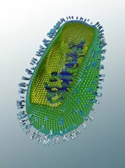
Influenza virus. Cut-away computer artwork of an influenza (flu) virus particle (virion). In each particles lipid envelope (blue) are two types of protein spike

Background imagePathogenic Collection: Influenza virus, artwork C018 / 2891

Influenza virus, artwork C018 / 2891
Influenza virus. Cut-away computer artwork of an influenza (flu) virus particle (virion). In each particles lipid envelope (blue) are two types of protein spike

Background imagePathogenic Collection: HIV particle, artwork C018 / 2895

HIV particle, artwork C018 / 2895
HIV particle, computer artwork. HIV (human immunodeficiency virus) causes AIDS (acquired immune deficiency syndrome). The virus consists of an RNA (ribonucleic acid) genome

Background imagePathogenic Collection: HIV particle, artwork C018 / 2900

HIV particle, artwork C018 / 2900
HIV particle, computer artwork. HIV (human immunodeficiency virus) causes AIDS (acquired immune deficiency syndrome). The virus consists of an RNA (ribonucleic acid) genome

Background imagePathogenic Collection: HIV particle, artwork C018 / 2896

HIV particle, artwork C018 / 2896
HIV particle, computer artwork. HIV (human immunodeficiency virus) causes AIDS (acquired immune deficiency syndrome). The virus consists of an RNA (ribonucleic acid) genome

Background imagePathogenic Collection: Influenza virus, artwork C018 / 2890

Influenza virus, artwork C018 / 2890
Influenza virus. Cut-away computer artwork of an influenza (flu) virus particle (virion). In each particles lipid envelope (green) are two types of protein spike

Background imagePathogenic Collection: Influenza virus, artwork C018 / 2892

Influenza virus, artwork C018 / 2892
Influenza virus. Cut-away computer artwork of an influenza (flu) virus particle (virion). In each particles lipid envelope (brown) are two types of protein spike

Background imagePathogenic Collection: Avian influenza virus, TEM C016 / 5843

Avian influenza virus, TEM C016 / 5843
Avian influenza virus, type A strain H7N9, coloured transmission electron micrograph (TEM). This virus first emerged in the human population in China, in March 2013

Background imagePathogenic Collection: Avian influenza virus, TEM C016 / 5841

Avian influenza virus, TEM C016 / 5841
Avian influenza virus, type A strain H7N9, coloured transmission electron micrograph (TEM). This virus first emerged in the human population in China, in March 2013

Background imagePathogenic Collection: Avian influenza virus, TEM C016 / 5842

Avian influenza virus, TEM C016 / 5842
Avian influenza virus, type A strain H7N9, coloured transmission electron micrograph (TEM). This virus first emerged in the human population in China, in March 2013

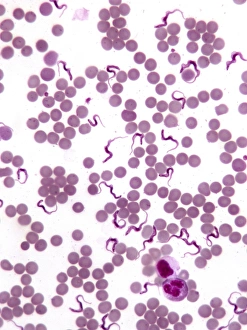
Background imagePathogenic Collection: Trypanosomes in blood smear, SEM C016 / 5783

Trypanosomes in blood smear, SEM C016 / 5783
Parasitic protozoan. Coloured scanning electron micrograph (SEM) of trypanosome protozoa (Trypanosoma sp.) in a blood smear

Background imagePathogenic Collection: Avian influenza virus, TEM C016 / 2354

Avian influenza virus, TEM C016 / 2354
Avian influenza virus, type A strain H7N9, coloured transmission electron micrograph (TEM). This virus first emerged in the human population in China, in March 2013

Background imagePathogenic Collection: Avian influenza virus, TEM C016 / 2353

Avian influenza virus, TEM C016 / 2353
Avian influenza virus, type A strain H7N9, coloured transmission electron micrograph (TEM). This virus first emerged in the human population in China, in March 2013

Background imagePathogenic Collection: Avian influenza virus, TEM C016 / 2352

Avian influenza virus, TEM C016 / 2352
Avian influenza virus, type A strain H7N9, coloured transmission electron micrograph (TEM). This virus first emerged in the human population in China, in March 2013

Background imagePathogenic Collection: Avian influenza virus, TEM C016 / 2351

Avian influenza virus, TEM C016 / 2351
Avian influenza virus, type A strain H7N9, coloured transmission electron micrograph (TEM). This virus first emerged in the human population in China, in March 2013

Background imagePathogenic Collection: Satellite tobacco necrosis virus C014 / 0005

Satellite tobacco necrosis virus C014 / 0005
Satellite tobacco necrosis virus. Computer artwork showing the surface structure of a tobacco necrosis satellivirus particle (virion)

Background imagePathogenic Collection: Hepatitis B virus particle C013 / 9983

Hepatitis B virus particle C013 / 9983
Hepatitis B virus particle. Computer artwork of the nucleocapsid of a hepatitis B virus (HBV) particle (virion). The capsid is a protein coat that encloses the viruss DNA (deoxyribonucleic acid)

Background imagePathogenic Collection: Canine parvovirus particle C013 / 9966

Canine parvovirus particle C013 / 9966
Canine parvovirus particle. Computer artwork showing the structure of the outer protein coat (capsid) of a canine parvovirus type 2 particle (virion)

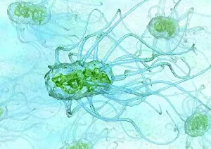
Background imagePathogenic Collection: Salmonella bacteria, artwork C013 / 7809

Salmonella bacteria, artwork C013 / 7809
Salmonella bacteria, computer artwork. Salmonella sp. bacteria are gram-negative rod-shaped bacteria that have flagella (hair-like structures) that they use for locomotion

Background imagePathogenic Collection: Salmonella bacteria, artwork C013 / 7808

Salmonella bacteria, artwork C013 / 7808
Salmonella bacteria, computer artwork. Salmonella sp. bacteria are gram-negative rod-shaped bacteria that have flagella (hair-like structures) that they use for locomotion

Background imagePathogenic Collection: Polyoma BK virus, artwork C013 / 7466

Polyoma BK virus, artwork C013 / 7466
Computer artwork of the capsid of a polyoma BK virus. This polyomavirus is common in the urinary tract of adults, where it lives without harming its host

Background imagePathogenic Collection: Bird flu virus particle, artwork C013 / 4650

Bird flu virus particle, artwork C013 / 4650
Bird flu virus particle. Computer artwork showing the internal and external structures of an influenza (flu) A subtype H5N1 (bird flu) virus particle (virion)

Background imagePathogenic Collection: Ross River virus particle, artwork C013 / 4641

Ross River virus particle, artwork C013 / 4641
Ross River virus particle. Computer artwork showing the outer structure of a Ross River virus (RRV) particle (virion). RRV is a small encapsulated single-strand RNA alphavirus endemic to Australia

Background imagePathogenic Collection: Marburg virus, artwork C013 / 4633

Marburg virus, artwork C013 / 4633
Marburg virus. Computer artwork of Marburg virus particles (virions). This tubular RNA (ribonucleic acid, coloured spheres) virus causes Marburg haemorrhagic fever in humans and non-human primates

Background imagePathogenic Collection: Hepatitis C virus, artwork

Hepatitis C virus, artwork
Hepatitis C virus. Computer artwork of hepatitis C virus particles (virions) in the human body. The virus consists of a core of RNA (ribonucleic acid) enclosed in a capsid

Background imagePathogenic Collection: EHEC E. coli bacteria, artwork C013 / 4621

EHEC E. coli bacteria, artwork C013 / 4621
EHEC E. coli bacteria. Computer artwork of a enterohaemorrhagic Escherichia coli (EHEC) bacteria in the human gut. E. coli bacteria are a normal part of the intestinal flora in humans

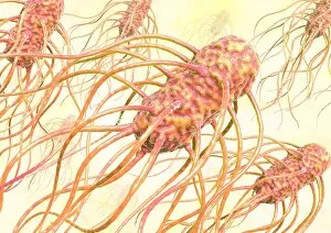
Background imagePathogenic Collection: Salmonella bacterium, artwork C013 / 4728

Salmonella bacterium, artwork C013 / 4728
Salmonella bacterium, computer artwork. Salmonella sp. bacteria are gram-negative rod-shaped bacteria that have flagella (hair-like structures) that they use for locomotion

Background imagePathogenic Collection: Salmonella bacteria, artwork C013 / 4727

Salmonella bacteria, artwork C013 / 4727
Salmonella bacteria, computer artwork. Salmonella sp. bacteria are gram-negative rod-shaped bacteria that have flagella (hair-like structures) that they use for locomotion

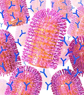
Background imagePathogenic Collection: Viral recognition by antibodies, artwork C013 / 4722

Viral recognition by antibodies, artwork C013 / 4722
Viral recognition by antibodies. Computer artwork of rabies (family Rhabdoviridae) virus particles (virions, pink) being identified by monoclonal antibodies (Y-shaped, blue)

Background imagePathogenic Collection: Rabies virus, artwork C013 / 4704

Rabies virus, artwork C013 / 4704
Rabies virus. Computer artwork of rabies (family Rhabdoviridae) virus particles (virions). The virus has a protein coat or capsid, which is made up of helically arranged subunits called capsomeres

Background imagePathogenic Collection: Rabies virus, artwork C013 / 4703

Rabies virus, artwork C013 / 4703
Rabies virus. Computer artwork of a rabies (family Rhabdoviridae) virus particle (virion). The virus has a protein coat or capsid, which is made up of helically arranged subunits called capsomeres

Background imagePathogenic Collection: Cabbage root infection, SEM

Cabbage root infection, SEM
Cabbage root infection. Coloured scanning electron micrograph (SEM) of a cross-section through one of the roots of a cabbage (Brassica sp)

Background imagePathogenic Collection: Erwinia bacteria, SEM

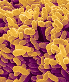
Erwinia bacteria, SEM
Erwinia bacteria. Coloured scanning electron micrograph (SEM) of Erwinia carotovora bacteria. This gram-negative flagellated bacterium causes soft rot of plant crops such as potato

Background imagePathogenic Collection: Grasshopper (Orthoptera sp. ) adult, killed by Pathogenic Fungus (Entomophthora sp)

Grasshopper (Orthoptera sp. ) adult, killed by Pathogenic Fungus (Entomophthora sp)
Grasshopper (Orthoptera sp.) adult, killed by Pathogenic Fungus (Entomophthora sp.) infection, fungus causes grasshopper to climb stalk before dying so spores will spread further, Pyrenees, Ariege

Background imagePathogenic Collection: Dance Fly (Empis tessellata) adult, fallen victim to Pathogenic Fungus (Entomophthora muscae)

Dance Fly (Empis tessellata) adult, fallen victim to Pathogenic Fungus (Entomophthora muscae) that enters brain and causes it to land, climb to the top of bush and die

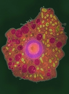
Background imagePathogenic Collection: Naegleria fowleri protozoa, TEM

Naegleria fowleri protozoa, TEM
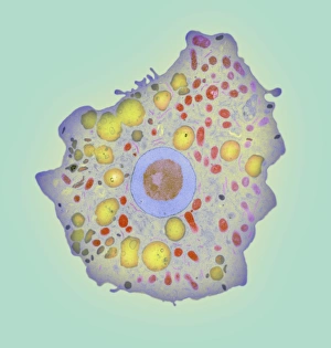
Naegleria fowleri protozoan. Coloured transmission electron micrograph (TEM) of a section through a Naegleria fowleri protozoan

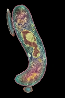
Background imagePathogenic Collection: Parasitic protozoan, TEM

Parasitic protozoan, TEM
Parasitic protozoan. Coloured transmission electron micrograph (TEM) of a trypanosome protozoan (Trypanosoma sp.). The nucleus (centre), cell membrane (outline)

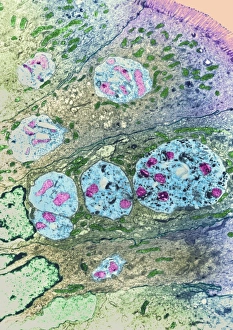
Background imagePathogenic Collection: Enterocytozoon sp. parasites, TEM

Enterocytozoon sp. parasites, TEM

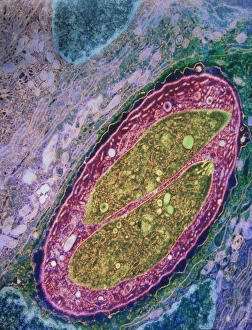
Background imagePathogenic Collection: Isospora sp. parasites, TEM

Isospora sp. parasites, TEM
Isospora sp. parasites. Coloured transmission electron micrograph (TEM) of Isospora sp. parasites (yellow) in a sectioned cell

Background imagePathogenic Collection: Naegleria fowleri protozoan, TEM

Naegleria fowleri protozoan, TEM
Naegleria fowleri protozoan. Coloured transmission electron micrograph (TEM) of a section through a Naegleria fowleri protozoan

Background imagePathogenic Collection: Hartmannella amoeba and bacterium

Hartmannella amoeba and bacterium. Computer artwork of a Hartmannella vermiformis amoeba (green) engulfing a Legionella pneumophila bacterium (orange) with a pseudopodium

Background imagePathogenic Collection: Nematode worms, SEM

Nematode worms, SEM
Nematode worm. Scanning electron micrograph of a juvenile Heterorhabditis bacteriophora nematode worm (centre) shedding its cuticle (outer coat, centre right) as it develops into an adult

Background imagePathogenic Collection: Lyme disease tick

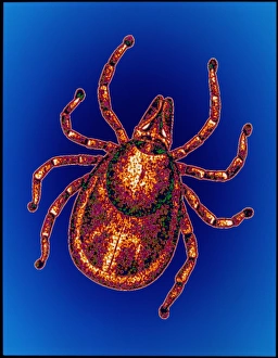
Lyme disease tick. Computer graphic illustration of the tick Ixodes ricinus, principal vector of Lyme disease in Europe. The head (above centre) contains a pair of palps

Background imagePathogenic Collection: Mosquito feeding

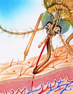
Mosquito feeding
Malaria mosquito. Illustration showing a mosquito on human skin, sucking blood. It has inserted its modified proboscis (red) into a superficial blood vessel


All Professionally Made to Order for Quick Shipping

Pathogenic microorganisms are microscopic entities that pose a significant threat to human health. From budding yeast cells to avian flu viruses, these tiny organisms have the potential to cause widespread diseases and infections. In the world of pathogens, the avian flu virus stands out as one of the most notorious culprits. Its ability to jump from birds to humans has sparked global concerns about pandemics and led scientists on a quest for effective prevention strategies. Neutrophils, our body's first line of defense against infection, play a crucial role in engulfing harmful bacteria like MRSA (methicillin-resistant Staphylococcus aureus). This SEM image captures this incredible process, showcasing how neutrophils work tirelessly to protect us from pathogenic invaders. Salmonella bacteria are another well-known pathogen responsible for causing foodborne illnesses worldwide. This SEM image provides an up-close look at their unique structure and highlights their capacity for survival in various environments. The HIV particle is infamous for its devastating impact on the immune system. Understanding its intricate structure has been instrumental in developing antiretroviral therapies that help manage this deadly virus. Anthrax cultures depicted in historical diagrams remind us of past outbreaks and highlight the importance of ongoing research into preventing bioterrorism threats associated with this highly infectious disease. Norovirus particles captured through TEM imaging reveal their distinctive shape and shed light on why they are so contagious. Responsible for many cases of gastroenteritis, noroviruses can spread rapidly within communities if proper hygiene measures aren't followed diligently. Tuberculosis bacteria continue to be a major global health concern due to their ability to infect millions each year. Efforts towards improved diagnostics and treatment options remain critical in combating this persistent pathogen. E. coli bacteria serve as both harmless inhabitants of our gut flora but also dangerous pathogens when certain strains acquire virulence factors. This diverse bacterium reminds us that not all E. coli strains are created equal.