Fraud Blocker Skip to main content

Pathogenic Collection (page 5)

Pathogenic microorganisms are microscopic entities that pose a significant threat to human health

Background imagePathogenic Collection: Eastern equine encephalitis, TEM C016 / 9390

Eastern equine encephalitis, TEM C016 / 9390
Eastern equine encephalitis. Transmission electron micrograph (TEM) of a section through tissue from a human central nervous system (CNS)

Background imagePathogenic Collection: MRSA bacteria, SEM C016 / 9422

MRSA bacteria, SEM C016 / 9422
MRSA bacteria. Coloured scanning electron micrograph (SEM) of methicillin-resistant Staphylococcus aureus (MRSA, round) bacteria

Background imagePathogenic Collection: Colorado tick fever virus infection, TEM C016 / 9384

Colorado tick fever virus infection, TEM C016 / 9384
Colorado tick fever virus infection. Transmission electron micrograph (TEM) of human tissue infected by Colorado tick fever virus (CTFV) particles (virions, dark circles)

Background imagePathogenic Collection: Syphilis bacterium, TEM C016 / 9461

Syphilis bacterium, TEM C016 / 9461
Syphilis bacteria. Transmission electron micrograph (TEM) of a Treponema pallidum spirochaete bacterium. T. pallidum is the bacteria that is responsible for the sexually transmitted infection syphilis

Background imagePathogenic Collection: Helicobacter pylori bacteria, SEM C016 / 9135

Helicobacter pylori bacteria, SEM C016 / 9135
Helicobacter pylori bacteria. Coloured scanning electron micrograph (SEM) of Helicobacter pylori bacteria (yellow) in the stomach. H

Background imagePathogenic Collection: Helicobacter pylori bacteria, SEM C016 / 9134

Helicobacter pylori bacteria, SEM C016 / 9134
Helicobacter pylori bacteria. Coloured scanning electron micrograph (SEM) of Helicobacter pylori bacteria (orange) in the stomach. H

Background imagePathogenic Collection: Swine flu virus particles, TEM C016 / 9399

Swine flu virus particles, TEM C016 / 9399
Swine flu virus particles. Transmission electron micrograph (TEM) of a section through H3N2 influenza A virus particles (virions)

Background imagePathogenic Collection: Swine flu virus particles, TEM C016 / 9406

Swine flu virus particles, TEM C016 / 9406
Influenza virus particles. Transmission electron micrograph (TEM) of influenza (flu) virus particles (virions). Each virion consists of ribonucleic acid (RNA, dark patches)

Background imagePathogenic Collection: Helicobacter pylori bacteria, SEM C016 / 9136

Helicobacter pylori bacteria, SEM C016 / 9136
Helicobacter pylori bacteria. Coloured scanning electron micrograph (SEM) of Helicobacter pylori bacteria (green) in the stomach. H

Background imagePathogenic Collection: Syphilis bacterium, TEM C016 / 9462

Syphilis bacterium, TEM C016 / 9462
Syphilis bacteria. Transmission electron micrograph (TEM) of a Treponema pallidum spirochaete bacterium. T. pallidum is the bacteria that is responsible for the sexually transmitted infection known

Background imagePathogenic Collection: St. Louis encephalitis virus particles C016 / 9453

St. Louis encephalitis virus particles C016 / 9453
St. Louis encephalitis virus particles. Coloured transmission electron micrograph (TEM) of a section through a mosquito salivary gland containing numerous Saint Louis encephalitis virus particles

Background imagePathogenic Collection: HIV virus particles, TEM C016 / 9404

HIV virus particles, TEM C016 / 9404
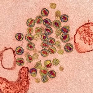
HIV virus particles. Transmission electron micrograph (TEM) of a section through human immunodeficiency virus (HIV) particles (virions, round)

Background imagePathogenic Collection: Smallpox virus particles, TEM C016 / 9447

Smallpox virus particles, TEM C016 / 9447
Smallpox virus particles. Coloured transmission electron micrograph (TEM) of a section through a tissue sample infected with numerous Variola virus particles (virions, brown)

Background imagePathogenic Collection: Spumavirus particles, TEM C016 / 9452

Spumavirus particles, TEM C016 / 9452
Spumavirus virus particles. Coloured transmission electron micrograph (TEM) of a section through a tissue sample infected with numerous spumavirus particles (virions, yellow)

Background imagePathogenic Collection: Varicella zoster virus particles, TEM C016 / 9466

Varicella zoster virus particles, TEM C016 / 9466
Varicella zoster virus particles. Transmission electron micrograph (TEM) of varicella zoster virus (VZV) particles (virions) from a patient with chickenpox

Background imagePathogenic Collection: Mycobacterium fortuitum bacteria C016 / 9423

Mycobacterium fortuitum bacteria C016 / 9423
Mycobacterium fortuitum bacteria, coloured scanning electron micrograph (SEM). These bacteria (yellow) are a non-tuberculosis strain of Mycobacterium

Background imagePathogenic Collection: Gonorrhoea bacteria, TEM C016 / 9425

Gonorrhoea bacteria, TEM C016 / 9425
Gonorrhoea bacteria. Transmission electron micrograph (TEM) of a diplococcal pair of Neisseria gonorrhoeae bacteria. N. gonorrhoeae is the bacteria that is responsible for the sexually transmitted

Background imagePathogenic Collection: HIV virus particles, TEM C016 / 9405

HIV virus particles, TEM C016 / 9405
HIV virus particles. Transmission electron micrograph (TEM) of a section through human immunodeficiency virus (HIV) particles (virions, round)

Background imagePathogenic Collection: White blood cell antigen presentation C016 / 9057

White blood cell antigen presentation C016 / 9057
White blood cell antigen presentation. Coloured scanning electron micrograph (SEM) showing the interaction between a macrophage (red) and a T helper lymphocyte (Th cell, yellow)

Background imagePathogenic Collection: Reovirus particles, TEM C016 / 9444

Reovirus particles, TEM C016 / 9444
Reovirus particles. Coloured transmission electron micrograph (TEM) of numerous reovirus particles (virions, blue). Reoviruses affect the gastrointestinal system and respiratory tract

Background imagePathogenic Collection: Swine flu virus particles, TEM C016 / 9400

Swine flu virus particles, TEM C016 / 9400
Swine flu virus particles. Transmission electron micrograph (TEM) of a section through H3N2 influenza A virus particles (virions)

Background imagePathogenic Collection: Orientia tsutsugamushi bacteria infection C016 / 9429

Orientia tsutsugamushi bacteria infection C016 / 9429
Orientia tsutsugamushi bacteria infection. Transmission electron micrograph (TEM) of a section through a capillary (red) infected with Orientia tsutsugamushi bacteria (blue). O

Background imagePathogenic Collection: MRSA bacteria, SEM C016 / 9421

MRSA bacteria, SEM C016 / 9421
MRSA bacteria. Coloured scanning electron micrograph (SEM) of methicillin-resistant Staphylococcus aureus (MRSA, round) bacteria

Background imagePathogenic Collection: Polio virus particles, TEM C016 / 9437

Polio virus particles, TEM C016 / 9437
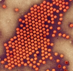
Poliovirus particles. Coloured transmission electron micrograph (TEM) of poliovirus particles (orange). Poliovirus infects children causing the disease poliomyelitis, or infantile paralysis

Background imagePathogenic Collection: Lassa virus particles, TEM C016 / 9408

Lassa virus particles, TEM C016 / 9408
Lassa virus particles. Transmission electron micrograph (TEM) of Lassa virus particles (virions, green) amongst cell debris. This Arenavirus is the cause of Lassa fever

Background imagePathogenic Collection: Legionella infected protozoan, TEM C016 / 9412

Legionella infected protozoan, TEM C016 / 9412
Legionella infected protozoan. Transmission electron micrograph (TEM) of a section through a single-celled Tetrahymena pyriformis protozoan, infected with Legionella pneumophila bacteria (yellow)

Background imagePathogenic Collection: Orientia tsutsugamushi bacterium C016 / 9430

Orientia tsutsugamushi bacterium C016 / 9430
Orientia tsutsugamushi bacterium. Transmission electron micrograph (TEM) of a section through an Orientia tsutsugamushi bacterium. O

Background imagePathogenic Collection: Ganjam virus infection, TEM C016 / 9396

Ganjam virus infection, TEM C016 / 9396
Ganjam virus infection. Transmission electron micrograph (TEM) of a section through human tissue infected with ganjam virus particles (virions, blue and yellow)

Background imagePathogenic Collection: Helicobacter pylori bacteria, SEM C016 / 9053

Helicobacter pylori bacteria, SEM C016 / 9053
Helicobacter pylori bacteria. Coloured scanning electron micrograph (SEM) of a Helicobacter pylori bacterium (blue) in the stomach. H

Background imagePathogenic Collection: HIV particles, artwork C016 / 9142

HIV particles, artwork C016 / 9142
HIV particles, computer artwork. HIV (human immunodeficiency virus) causes AIDS (acquired immune deficiency syndrome). The virus consists of an RNA (ribonucleic acid) genome (pink)

Background imagePathogenic Collection: Swine flu virus particles, TEM C016 / 9407

Swine flu virus particles, TEM C016 / 9407
Influenza virus particles. Transmission electron micrograph (TEM) of influenza (flu) virus particles (virions). Each virion consists of ribonucleic acid (RNA, dark patches)

Background imagePathogenic Collection: Giardia protozoan, SEM C016 / 9398

Giardia protozoan, SEM C016 / 9398
Giardia protozoan. Coloured scanning electron micrograph (SEM) of a Giardia sp. trophozoite protozoan (yellow) attached to the lining of a human intestine

Background imagePathogenic Collection: Varicella zoster virus particles, TEM C016 / 9467

Varicella zoster virus particles, TEM C016 / 9467
Varicella zoster virus particles. Transmission electron micrograph (TEM) of varicella zoster virus (VZV) particles (virions) from a patient with chickenpox

Background imagePathogenic Collection: Meningitis bacteria, SEM

Meningitis bacteria, SEM
Meningitis bacteria. Coloured scanning electron micrograph (SEM) of Neisseria meningitidis bacteria. N. meningitidis is an obligate human parasite that causes meningococcal meningitis

Background imagePathogenic Collection: Flu virus, TEM

Flu virus, TEM
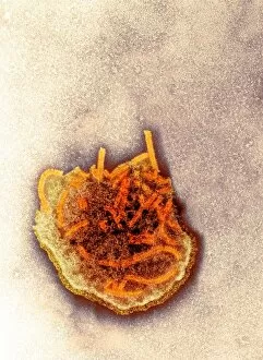
Flu virus. Transmission electron micrograph (TEM) of a section through influenza (flu) virus particles (virions, purple) budding from a host cell. This is the final stage in viral replication

Background imagePathogenic Collection: Sleeping sickness parasites, SEM

Sleeping sickness parasites, SEM
Sleeping sickness parasites. Coloured scanning electron micrograph (SEM) of a Trypanosoma brucei parasites (blue) and white blood cells (pink) in a mouse liver. T
Background imagePathogenic Collection: Sleeping sickness parasite, SEM

Sleeping sickness parasite, SEM
Sleeping sickness parasite. Coloured scanning electron micrograph (SEM) of a Trypanosoma brucei parasite (blue) and a white blood cell (green) in a mouse liver. T

Background imagePathogenic Collection: Drug effect on viruses, conceptual image C016 / 6253

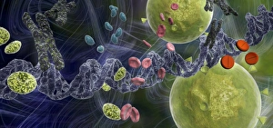
Drug effect on viruses, conceptual image C016 / 6253
Drug effect on viruses, conceptual image. Computer artwork showing a single strand of DNA (deoxyribonucleic acid, spiral, centre), red blood cells (pink), virus particles (virions, green, small)

Background imagePathogenic Collection: Measles virus particle, TEM C015 / 7160

Measles virus particle, TEM C015 / 7160
Measles virus particle, coloured transmission electron micrograph (TEM). This virus, from the Morbillivirus group of viruses, consists of an RNA (ribonucleic acid)

Background imagePathogenic Collection: Measles virus particle, TEM C015 / 7161

Measles virus particle, TEM C015 / 7161
Measles virus particle, coloured transmission electron micrograph (TEM). This virus, from the Morbillivirus group of viruses, consists of an RNA (ribonucleic acid)

Background imagePathogenic Collection: Bird flu research C015 / 8803

Bird flu research C015 / 8803
Bird flu research. Researcher harvesting influenza type a H7N9 viruses from cultures grown in eggs. This virus first emerged in the human population in China, in March 2013

Background imagePathogenic Collection: Avian influenza virus, TEM C015 / 8800

Avian influenza virus, TEM C015 / 8800
Avian influenza virus, type A strain H7N9, coloured transmission electron micrograph (TEM). This virus first emerged in the human population in China, in March 2013

Background imagePathogenic Collection: Measles virus particle, TEM C015 / 7162

Measles virus particle, TEM C015 / 7162
Measles virus particle, coloured transmission electron micrograph (TEM). This virus, from the Morbillivirus group of viruses

Background imagePathogenic Collection: Bird flu diagnostic kit C015 / 8805

Bird flu diagnostic kit C015 / 8805
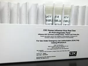
Bird flu diagnostic kit. Reagents for diagnosing influenza type a H7N9 infection in humans. This virus first emerged in the human population in China, in March 2013

Background imagePathogenic Collection: Measles virus particle, TEM C015 / 7159

Measles virus particle, TEM C015 / 7159
Measles virus particle, coloured transmission electron micrograph (TEM). This virus, from the Morbillivirus group of viruses, consists of an RNA (ribonucleic acid)

Background imagePathogenic Collection: Avian influenza virus, TEM C015 / 8799

Avian influenza virus, TEM C015 / 8799
Avian influenza virus, type A strain H7N9, coloured transmission electron micrograph (TEM). This virus first emerged in the human population in China, in March 2013

Background imagePathogenic Collection: Bird flu research C015 / 8804

Bird flu research C015 / 8804
Bird flu research. Researcher measuring harvested influenza type a H7N9 viruses grown in the lab. This virus first emerged in the human population in China, in March 2013

Background imagePathogenic Collection: Avian influenza virus, TEM C015 / 8797

Avian influenza virus, TEM C015 / 8797
Avian influenza virus, type A strain H7N9, coloured transmission electron micrograph (TEM). This virus first emerged in the human population in China, in March 2013


All Professionally Made to Order for Quick Shipping

Pathogenic microorganisms are microscopic entities that pose a significant threat to human health. From budding yeast cells to avian flu viruses, these tiny organisms have the potential to cause widespread diseases and infections. In the world of pathogens, the avian flu virus stands out as one of the most notorious culprits. Its ability to jump from birds to humans has sparked global concerns about pandemics and led scientists on a quest for effective prevention strategies. Neutrophils, our body's first line of defense against infection, play a crucial role in engulfing harmful bacteria like MRSA (methicillin-resistant Staphylococcus aureus). This SEM image captures this incredible process, showcasing how neutrophils work tirelessly to protect us from pathogenic invaders. Salmonella bacteria are another well-known pathogen responsible for causing foodborne illnesses worldwide. This SEM image provides an up-close look at their unique structure and highlights their capacity for survival in various environments. The HIV particle is infamous for its devastating impact on the immune system. Understanding its intricate structure has been instrumental in developing antiretroviral therapies that help manage this deadly virus. Anthrax cultures depicted in historical diagrams remind us of past outbreaks and highlight the importance of ongoing research into preventing bioterrorism threats associated with this highly infectious disease. Norovirus particles captured through TEM imaging reveal their distinctive shape and shed light on why they are so contagious. Responsible for many cases of gastroenteritis, noroviruses can spread rapidly within communities if proper hygiene measures aren't followed diligently. Tuberculosis bacteria continue to be a major global health concern due to their ability to infect millions each year. Efforts towards improved diagnostics and treatment options remain critical in combating this persistent pathogen. E. coli bacteria serve as both harmless inhabitants of our gut flora but also dangerous pathogens when certain strains acquire virulence factors. This diverse bacterium reminds us that not all E. coli strains are created equal.