
Patellar Ligament Collection
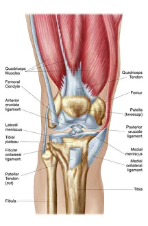
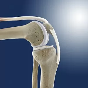
The patellar ligament, also known as the patellar tendon, is a crucial component of the human knee joint


All Professionally Made to Order for Quick Shipping
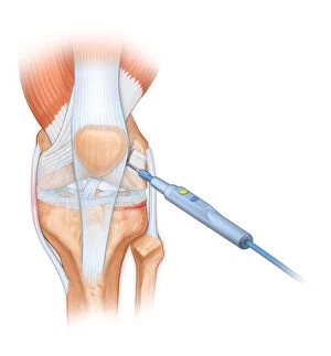
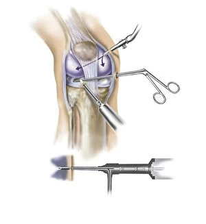
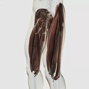
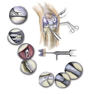
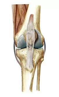
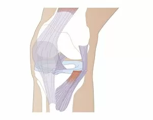
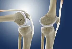
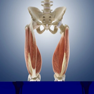
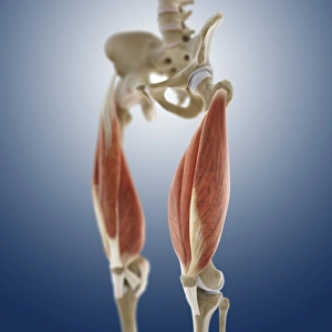
The patellar ligament, also known as the patellar tendon, is a crucial component of the human knee joint. It plays a significant role in stabilizing and supporting the knee during various movements and activities. In an intricate anatomy of the human knee joint, the patellar ligament stands out as a strong band that connects the kneecap (patella) to the tibia bone. This connection allows for efficient transmission of forces from the thigh muscles to the lower leg, enabling smooth movement. During certain surgical procedures like using a bovie to cut through retinaculum or cleaning up femur of displaced patellar knee, precise attention is given to this ligament. Its integrity must be maintained while addressing any issues related to it or surrounding structures. A detailed illustration of a human knee cutaway reveals how arthroscopic instruments are inserted into specific areas around the insertion point of this vital ligament. This technique ensures minimal invasiveness while performing necessary interventions. From both side and anterior views of male muscle anatomy in human legs, one can observe how these powerful muscles interact with and influence the function of not only the patellar ligament but also other components within this complex joint system. As part of typical synovial joints found in our body, such as knees, elbows, and shoulders; understanding its structure becomes essential for medical professionals conducting arthroscopic surgical procedures on patients' knees. The detail-oriented approach helps them navigate through different layers without compromising patient safety or desired outcomes.