Fraud Blocker Skip to main content

Parasitic Collection (page 10)

"Exploring the Intricate World Creatures: From Gastrointestinal Nematodes to Eyelash Mites" Delving into the hidden realm of parasites

Background imageParasitic Collection: Cat fluke, light micrograph C014 / 4858

Cat fluke, light micrograph C014 / 4858
Cat fluke (Opisthorchis felineus), light micrograph. This parasitic trematode (flatworm) infests the liver of members of the cat family

Background imageParasitic Collection: Cat flea head, SEM C014 / 4842

Cat flea head, SEM C014 / 4842
Cat flea head. Coloured scanning electron micrograph (SEM) of the head of cat flea (Ctenocephalides felis). Its eyes (red) can be seen at either side of its head

Background imageParasitic Collection: Tapeworm head, SEM C014 / 4877

Tapeworm head, SEM C014 / 4877
Tapeworm head. Coloured scanning electron micrograph of the head (scolex) of a tapeworm (class Cestoda) found in a shark, showing the hooks used to attach to the host animals digestive tract

Background imageParasitic Collection: Tapeworm head, SEM C014 / 4880

Tapeworm head, SEM C014 / 4880
Tapeworm head. Coloured scanning electron micrograph of the head (scolex) of a tapeworm (Platybothrium auriculatum) found in a blue shark (Prionace glauca)

Background imageParasitic Collection: Beelouse, light micrograph C014 / 4841

Beelouse, light micrograph C014 / 4841
Beelouse (Braula coeca), dark-field illuminated light micrograph. This wingless fly is a parasite of the honeybee (Apis mellifera)

Background imageParasitic Collection: Parasitic louse, light micrograph C014 / 4850

Parasitic louse, light micrograph C014 / 4850
Parasitic louse (Haematopinus asini), light micrograph. This louse commonly infects horses. It causes irritation that can cause damage by the horse rubbing itself raw

Background imageParasitic Collection: Paraneplocephala tapeworm, micrograph C014 / 4859

Paraneplocephala tapeworm, micrograph C014 / 4859
Paraneplocephala tapeworm. Light micrograph of a Paraneplocephala sp. tapeworm. This short parasitic tapeworm (order Cestoidea) is found in horses and other herbivores

Background imageParasitic Collection: Strepsipteran parasitic insect, SEM C014 / 4876

Strepsipteran parasitic insect, SEM C014 / 4876
Strepsipteran parasitic insect. Coloured scanning electron micrograph (SEM) of the head of a strepsipteran. The strepsipterans are an unusual parasitic group of insects that are distantly related to

Background imageParasitic Collection: Tapeworm head, SEM C014 / 4878

Tapeworm head, SEM C014 / 4878
Tapeworm head. Coloured scanning electron micrograph of the head (scolex) of a tapeworm (class Cestoda) found in a shark, showing the hooks used to attach to the host animals digestive tract

Background imageParasitic Collection: Strepsipteran parasitic insect, SEM C014 / 4875

Strepsipteran parasitic insect, SEM C014 / 4875
Strepsipteran parasitic insect, coloured scanning electron micrograph (SEM). The strepsipterans are an unusual parasitic group of insects that are distantly related to beetles

Background imageParasitic Collection: Strepsipteran parasitic insect, SEM C014 / 4874

Strepsipteran parasitic insect, SEM C014 / 4874
Strepsipteran parasitic insect, coloured scanning electron micrograph (SEM). The strepsipterans are an unusual parasitic group of insects that are distantly related to beetles

Background imageParasitic Collection: Paraneplocephala tapeworm, micrograph C014 / 4860

Paraneplocephala tapeworm, micrograph C014 / 4860
Paraneplocephala tapeworm. Light micrograph showing the internal anatomy of a Paraneplocephala sp. tapeworm. This short parasitic tapeworm (order Cestoidea) is found in horses and other herbivores

Background imageParasitic Collection: Schistosome flukes mating, micrograph C014 / 4867

Schistosome flukes mating, micrograph C014 / 4867
Schistosome flukes mating. Light micrograph of Schistosoma japonicum fluke worms mating. The male is the smaller animal at centre

Background imageParasitic Collection: Tapeworm head, SEM C014 / 4879

Tapeworm head, SEM C014 / 4879
Tapeworm head. Coloured scanning electron micrograph of the head (scolex) of a tapeworm (class Cestoda) found in a shark, showing the suckers used to attach to the host animals digestive tract

Background imageParasitic Collection: Common liver fluke C014 / 4847

Common liver fluke C014 / 4847
Common liver fluke (Fasciola hepatica). This parasitic trematode (flatworm) infects the livers of various mammals, including humans

Background imageParasitic Collection: Beaver beetle leg, SEM

Beaver beetle leg, SEM
Beaver beetle leg. Coloured scanning electron micrograph (SEM) of a close-up on the fore-leg of a beaver (Platypsyllus sp.) beetle

Background imageParasitic Collection: Honey bee mite, SEM C016 / 8024

Honey bee mite, SEM C016 / 8024
Honey bee mite. Coloured scanning electron micrograph (SEM) of the underside of a Varroa sp. mite from a honey bee. Varroa are parasitic mites that attack honey bees (Apis sp.)

Background imageParasitic Collection: Cicada infected with fungus C014 / 4581

Cicada infected with fungus C014 / 4581
Cicada infected with fungus. Close-up of a dead cicada (family Cicadidae) that has been killed by a parasitic fungus

Background imageParasitic Collection: Cicada infected with fungus C014 / 4582

Cicada infected with fungus C014 / 4582
Cicada infected with fungus. Dead cicada (family Cicadidae) that has been killed by a parasitic fungus

Background imageParasitic Collection: Cicada infected with fungus C014 / 4580

Cicada infected with fungus C014 / 4580
Cicada infected with fungus. Dead cicada (family Cicadidae) that has been killed by a parasitic fungus

Background imageParasitic Collection: Common spangle galls on a leaf

Common spangle galls on a leaf
Common spangle galls on the underside of an oak (Quercus sp.) leaf. These protective galls contain the larvae of a parasitic wasp (Neuroterus quercusbaccarum). Photographed in Poland

Background imageParasitic Collection: Fungus parasitising a fly

Fungus parasitising a fly. Close-up of a fly that has been killed by the parasitic fungus Entomophthora muscae. This fungus is highly adapted to its host

Background imageParasitic Collection: Silk button spangle galls on a leaf

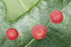
Silk button spangle galls on a leaf
Silk button spangle galls on the underside of an oak (Quercus sp.) leaf. These protective galls contain the larvae of a parasitic wasp (Neuroterus numismalis). Photographed in Poland

Background imageParasitic Collection: Red Broomrape (Hyobanche sanguinea)

Red Broomrape (Hyobanche sanguinea) in flower. Photographed in Cape area, South Africa

Background imageParasitic Collection: Malarial blood cell, SEM C017 / 8307

Malarial blood cell, SEM C017 / 8307
Malarial blood cell. Coloured scanning electron micrograph (SEM) of an erythrocyte (red blood cell) infected with Plasmodium parasites, the cause of malaria

Background imageParasitic Collection: Parasitic shell on starfish

Parasitic shell on starfish
A parasitic shell, Thyca crystallina, growing on its host starfish. The shell only attaches to this type of starfish, burrows into the it and sucks up nutrients

Background imageParasitic Collection: Fungus on a redwood tree C018 / 2574

Fungus on a redwood tree C018 / 2574
Fungus on a redwood tree. Tree fungus growing on a coast redwood (Sequoia sempervirens) tree. Photographed in Muir Woods National Park, California, USA

Background imageParasitic Collection: Nurse with a jar of medicinal leeches C018 / 2317

Nurse with a jar of medicinal leeches C018 / 2317
Medicinal leeches (Hirudo medicinalis) in a tank of water. Leeches are parasites that feed on blood. They attach themselves to the skin using suckers and create a wound with three sharp jaw plates

Background imageParasitic Collection: Dodder parasitising a tomato plant

Dodder parasitising a tomato plant. Dodder (Cuscuta sp.) tendril (yellow) wrapping itself around a tomato (Solanum lycopersicum) plant

Background imageParasitic Collection: Blood flukes mating C016 / 5782

Blood flukes mating C016 / 5782
Blood flukes (Schistosoma sp.) mating. These parasites are found in man and other mammals

Background imageParasitic Collection: Horse botfly larvae C016 / 5712

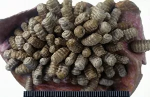
Horse botfly larvae C016 / 5712
Horse botfly (Gasterophilus sp.). Specimen of hose botfly larvae attached to the stomach wall of a horse

Background imageParasitic Collection: Conopid fly C016 / 4714

Conopid fly C016 / 4714
Conopid fly (Myopa buccata) on a plant. Conopid flies (family Conopidae) are common throughout much of Europe. Their larvae are endoparasites of bumble bees (Bombus sp.). Photographed in the UK

Background imageParasitic Collection: Chalcid wasp laying eggs in an oak gall C016 / 4710

Chalcid wasp laying eggs in an oak gall C016 / 4710
Chalcid wasp laying eggs in an oak gall. Close-up of a chalcid wasp (family Torymidae) hyperparasitising oak apple galls of the gall wasp (Biorhiza pallida)

Background imageParasitic Collection: Zombie ant fungus life-cycle, artwork

Zombie ant fungus life-cycle, artwork
Zombie ant fungus life-cycle. Computer artwork showing the life cycle of the parasitoidal fungus Ophiocordyceps unilateralis

Background imageParasitic Collection: Oak Mazegill (Daedalea quercina) C016 / 3601

Oak Mazegill (Daedalea quercina) C016 / 3601
Oak Mazegill (Daedalea quercina) growing on an oak log in autumn. Photographed in Exmoor National Park, Devon

Background imageParasitic Collection: Ivy Broomrape (Orobanche hederae) C016 / 3485

Ivy Broomrape (Orobanche hederae) C016 / 3485
Ivy Broomrape (Orobanche hederae) in flower. This plant is parasitic on ivy (Hedera sp.). Photographed in Picos de Europa, Spain

Background imageParasitic Collection: Ivy Broomrape (Orobanche hederae) C016 / 3484

Ivy Broomrape (Orobanche hederae) C016 / 3484
Ivy Broomrape (Orobanche hederae) in flower. This plant is parasitic on ivy (Hedera sp.). Photographed in Picos de Europa, Spain

Background imageParasitic Collection: Slender Broomrape (Orobanche gracilis) C016 / 3483

Slender Broomrape (Orobanche gracilis) C016 / 3483
Slender Broomrape (Orobanche gracilis) in flower. This plant is parasitic on legumes. Photographed in Picos de Europa, Spain

Background imageParasitic Collection: Tapeworm head, SEM C016 / 3093

Tapeworm head, SEM C016 / 3093
Tapeworm head. Coloured scanning electron micrograph (SEM) of the head of a tapeworm (class Cestoda) in the cysticercoid stage

Background imageParasitic Collection: Tapeworm head, SEM C016 / 3092

Tapeworm head, SEM C016 / 3092
Tapeworm head. Coloured scanning electron micrograph (SEM) of the head of a tapeworm (class Cestoda) in the cysticercoid stage

Background imageParasitic Collection: Gapeworm, SEM C016 / 3085

Gapeworm, SEM C016 / 3085
Gapeworm. Coloured scanning electron micrograph of a female gapeworm (Syngamus trachea) from an owl. Gapeworms are parasitic nematode worms that infect the trachea (windpipe) of certain birds

Background imageParasitic Collection: Gapeworm, SEM C016 / 3084

Gapeworm, SEM C016 / 3084
Gapeworm. Coloured scanning electron micrograph of a female gapeworm (Syngamus trachea) from an owl. Gapeworms are parasitic nematode worms that infect the trachea (windpipe) of certain birds

Background imageParasitic Collection: Gapeworm head, SEM C016 / 3082

Gapeworm head, SEM C016 / 3082
Gapeworm head. Coloured scanning electron micrograph of the head of a female gapeworm (Syngamus trachea) from an owl. Gapeworms are parasitic nematode worms that infect the trachea (windpipe)

Background imageParasitic Collection: Gapeworm head, SEM C016 / 3083

Gapeworm head, SEM C016 / 3083
Gapeworm head. Coloured scanning electron micrograph of the head of a female gapeworm (Syngamus trachea) from an owl. Gapeworms are parasitic nematode worms that infect the trachea (windpipe)

Background imageParasitic Collection: Malaria parasite in red blood cell C016 / 2444

Malaria parasite in red blood cell C016 / 2444
Malaria parasite in red blood cell. Light micrograph of a red blood cell (erythrocyte) infected with a Plasmodim vivax protozoan. This parasite causes recurring malaria in humans

Background imageParasitic Collection: Malaria parasite in red blood cell C016 / 2443

Malaria parasite in red blood cell C016 / 2443
Malaria parasite in red blood cell. Light micrograph of a red blood cell (erythrocyte) infected with a Plasmodim vivax protozoan. This parasite causes recurring malaria in humans

Background imageParasitic Collection: Malaria parasite in red blood cell C016 / 2442

Malaria parasite in red blood cell C016 / 2442
Malaria parasite in red blood cell. Light micrograph of a red blood cell (erythrocyte) infected with a Plasmodim vivax protozoan. This parasite causes recurring malaria in humans

Background imageParasitic Collection: Schistosome fluke worms, artwork C013 / 4642

Schistosome fluke worms, artwork C013 / 4642
Schistosome fluke worms. Computer artwork of Schistosoma mansoni fluke (trematode) worms in the human body. S. mansoni live in the veins around the large intestine and cause schistosomiasis


All Professionally Made to Order for Quick Shipping

"Exploring the Intricate World Creatures: From Gastrointestinal Nematodes to Eyelash Mites" Delving into the hidden realm of parasites, we encounter gastrointestinal nematodes, microscopic worms that reside in the intestines of various animals. Behold the intricate head structure of a dog tapeworm under scanning electron microscopy (SEM), revealing its remarkable adaptation for survival within its host. Zooming in even closer, SEM unveils the minuscule world of a head louse, showcasing its gripping claws and needle-like mouthparts used for feeding on human blood. Meet the sheep tick, an arachnid parasite found in grassy landscapes; SEM allows us to appreciate its formidable appendages designed for clinging onto hosts like sheep or deer. In our own eyelashes lies an unexpected inhabitant – the eyelash mite. SEM captures their tiny bodies and long tails as they navigate through this often overlooked ecosystem. Journeying deep into Sri Lanka's lush jungles reveals a diverse array organisms coexisting with their hosts amidst nature's beauty and complexity. Witnessing a female mosquito's internal anatomy while sucking blood from human skin through cross-section imagery sheds light on these pesky yet fascinating disease vectors. An illustrated depiction showcases the life cycle and morphology of tapeworms, highlighting their ability to adapt across different host species throughout their complex lifecycle. Examining eyelash mite tails under SEM unravels further mysteries about these elusive creatures that call our lashes home – truly captivating microcosms within ourselves. Venturing underwater brings us face-to-face with sea lampreys; whether it be Petromyzon marinus or other species like lamperns and silver lampreys - these jawless fish are notorious parasites known for attaching themselves to larger marine animals.