Fraud Blocker Skip to main content

Parasitic Collection (page 9)

"Exploring the Intricate World Creatures: From Gastrointestinal Nematodes to Eyelash Mites" Delving into the hidden realm of parasites

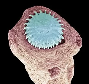
Background imageParasitic Collection: Dog tapeworm, SEM C016 / 9039

Dog tapeworm, SEM C016 / 9039
Dog tapeworm. Coloured scanning electron micrograph (SEM) of a dog tapeworm (Echinococcus granulosus). Tapeworms are parasitic flatworms that live in the digestive tract of their vertebrate host. E

Background imageParasitic Collection: Beef tapeworm, SEM C016 / 9079

Beef tapeworm, SEM C016 / 9079
Beef tapeworm. Coloured scanning electron micrograph (SEM) of the head of a beef tapeworm (Taenia saginata) showing the suckers it uses to cling to its host

Background imageParasitic Collection: Beef tapeworm, SEM C016 / 9078

Beef tapeworm, SEM C016 / 9078
Beef tapeworm. Coloured scanning electron micrograph (SEM) of the head of a beef tapeworm (Taenia saginata) showing the suckers it uses to cling to its host

Background imageParasitic Collection: Flea tapeworm, SEM C016 / 9034

Flea tapeworm, SEM C016 / 9034
Flea tapeworm. Coloured scanning electron micrograph (SEM) of a flea tapeworm (Dipylidium caninum), showing its head (top)

Background imageParasitic Collection: Flea tapeworm, SEM C016 / 9035

Flea tapeworm, SEM C016 / 9035
Flea tapeworm. Coloured scanning electron micrograph (SEM) of a flea tapeworm (Dipylidium caninum), showing its head (top)

Background imageParasitic Collection: Tapeworm, SEM C016 / 9080

Tapeworm, SEM C016 / 9080
Tapeworm. Coloured scanning electron micrograph (SEM) of the head of a tapeworm (Taenia pisiformis), showing the hooks (centre) it uses to cling to its host

Background imageParasitic Collection: Tapeworm, SEM C016 / 9077

Tapeworm, SEM C016 / 9077
Tapeworm. Coloured scanning electron micrograph (SEM) of a tapeworm (Taenia pisiformis), showing the hooks (upper right) it uses to cling to its host

Background imageParasitic Collection: Tapeworm, SEM C016 / 9081

Tapeworm, SEM C016 / 9081
Tapeworm. Coloured scanning electron micrograph (SEM) of the head of a tapeworm (Taenia pisiformis), showing the hooks (centre) it uses to cling to its host

Background imageParasitic Collection: Pork tapeworm, SEM C016 / 9071

Pork tapeworm, SEM C016 / 9071
Pork tapeworm. Coloured scanning electron micrograph (SEM) of the head of a pork tapeworm (Taenia solium), showing the hooks it uses to cling to its host

Background imageParasitic Collection: Tapeworm, SEM C016 / 9076

Tapeworm, SEM C016 / 9076
Tapeworm. Coloured scanning electron micrograph (SEM) of a tapeworm (Taenia pisiformis), showing the hooks (upper right) it uses to cling to its host

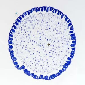
Background imageParasitic Collection: Roundworm germ cells, light micrograph C016 / 9538

Roundworm germ cells, light micrograph C016 / 9538
Roundworm germ cells. Light micrograph of germ cells from a roundworm (Ascaris sp.), undergoing mitosis (nuclear division)

Background imageParasitic Collection: Dog tapeworm, SEM C016 / 9038

Dog tapeworm, SEM C016 / 9038
Dog tapeworm. Coloured scanning electron micrograph (SEM) of a dog tapeworm (Echinococcus granulosus). Tapeworms are parasitic flatworms that live in the digestive tract of their vertebrate host. E

Background imageParasitic Collection: Pork tapeworm, SEM C016 / 9072

Pork tapeworm, SEM C016 / 9072
Pork tapeworm. Coloured scanning electron micrograph (SEM) of the head of a pork tapeworm (Taenia solium), showing the hooks it uses to cling to its host

Background imageParasitic Collection: Hartmannella vermiformis protozoa cysts C016 / 9402

Hartmannella vermiformis protozoa cysts C016 / 9402
Hartmannella vermiformis protozoa cysts. Transmission electron micrograph (TEM) of a section through cysts (round) containing Hartmannella vermiformis protozoa. H

Background imageParasitic Collection: Legionella infected protozoan, TEM C016 / 9412

Legionella infected protozoan, TEM C016 / 9412
Legionella infected protozoan. Transmission electron micrograph (TEM) of a section through a single-celled Tetrahymena pyriformis protozoan, infected with Legionella pneumophila bacteria (yellow)

Background imageParasitic Collection: Dog tapeworm, SEM C016 / 9036

Dog tapeworm, SEM C016 / 9036
Dog tapeworm. Coloured scanning electron micrograph (SEM) of the head of a dog tapeworm (Echinococcus granulosus), showing the hooks it uses to cling to its host

Background imageParasitic Collection: Dog tapeworm, SEM C016 / 9037

Dog tapeworm, SEM C016 / 9037
Dog tapeworm. Coloured scanning electron micrograph (SEM) of the head of a dog tapeworm (Echinococcus granulosus), showing the hooks it uses to cling to its host

Background imageParasitic Collection: Giardia protozoan, SEM C016 / 9398

Giardia protozoan, SEM C016 / 9398
Giardia protozoan. Coloured scanning electron micrograph (SEM) of a Giardia sp. trophozoite protozoan (yellow) attached to the lining of a human intestine

Background imageParasitic Collection: Beef tapeworm, SEM C016 / 9075

Beef tapeworm, SEM C016 / 9075
Beef tapeworm. Coloured scanning electron micrograph (SEM) of the head of a beef tapeworm (Taenia saginata) showing the suckers it uses to cling to its host

Background imageParasitic Collection: Pork tapeworm, SEM C016 / 9073

Pork tapeworm, SEM C016 / 9073
Pork tapeworm. Coloured scanning electron micrograph (SEM) of the head of a pork tapeworm (Taenia solium), showing the hooks it uses to cling to its host

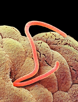
Background imageParasitic Collection: Threadworms in the gut, SEM

Threadworms in the gut, SEM
Threadworms in the gut. Coloured scanning electron micrograph (SEM) showing threadworms (Enterobius sp. orange) on the interior surface of a human intestine

Background imageParasitic Collection: Sleeping sickness parasites, SEM

Sleeping sickness parasites, SEM
Sleeping sickness parasites. Coloured scanning electron micrograph (SEM) of a Trypanosoma brucei parasites (blue) and white blood cells (pink) in a mouse liver. T

Background imageParasitic Collection: Sleeping sickness parasite, SEM

Sleeping sickness parasite, SEM
Sleeping sickness parasite. Coloured scanning electron micrograph (SEM) of a Trypanosoma brucei parasite (blue) and a white blood cell (green) in a mouse liver. T

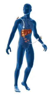
Background imageParasitic Collection: Malaria infection cycle, artwork

Malaria infection cycle, artwork. Life cycle of the Plasmodium parasite that causes malaria, which can cause coma and death. At upper left, a female Anopheles mosquito bites and infects a person

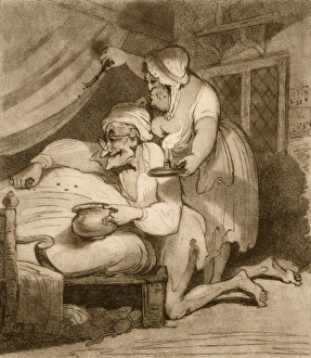
Background imageParasitic Collection: Catching bedbugs, 18th century

Catching bedbugs, 18th century
Catching bedbugs. 18th-century artwork, titled Summer Amusement - Bugg Hunting, showing an elderly couple catching bedbugs

Background imageParasitic Collection: Rust fungus on a bellflower leaf, SEM C019 / 0239

Rust fungus on a bellflower leaf, SEM C019 / 0239
Rust fungus. Coloured scanning electron micrograph (SEM) of a rust fungus (order Pucciniales) on a bellflower (Campanula sp.) plant

Background imageParasitic Collection: Rust fungus on a bellflower leaf, SEM C019 / 0238

Rust fungus on a bellflower leaf, SEM C019 / 0238
Rust fungus. Coloured scanning electron micrograph (SEM) of a rust fungus (order Pucciniales) on a bellflower (Campanula sp.) plant

Background imageParasitic Collection: Scale insect, SEM C019 / 0305

Scale insect, SEM C019 / 0305
Scale insect. Coloured scanning electron micrograph (SEM) of a scale insect (superfamily Coccoidea) on a leaf. This pest feeds on the plants sap

Background imageParasitic Collection: Scale insect, SEM C019 / 0306

Scale insect, SEM C019 / 0306
Scale insect. Coloured scanning electron micrograph (SEM) of a scale insect (superfamily Coccoidea) on a leaf. This pest feeds on the plants sap

Background imageParasitic Collection: Scale insects C014 / 2575

Scale insects C014 / 2575
Scale insects (superfamily Coccoidea) on a plant. This pest feeds on the plants sap. It secretes a powdery wax coating that protects it against pesticides and predators

Background imageParasitic Collection: Palo Verde tree and mistletoe C014 / 1546

Palo Verde tree and mistletoe C014 / 1546
Palo Verde tree and mistletoe. Blue Palo Verde (Parkinsonia florida) tree with desert mistletoe (Phoradendron californicum, dark) growing in its branches

Background imageParasitic Collection: Tick on a snake C014 / 0957

Tick on a snake C014 / 0957
Tick on a snake. Parasitic tick (superfamily Ixodoidea, white) embedded in the body of Waglers sipo, or smooth machete savane, (Chironius scurrulus) snake

Background imageParasitic Collection: Palo Verde tree and mistletoe C014 / 1548

Palo Verde tree and mistletoe C014 / 1548
Palo Verde tree and mistletoe. Blue Palo Verde (Parkinsonia florida, left) tree with desert mistletoe (Phoradendron californicum, dark) growing in its branches

Background imageParasitic Collection: Parasitic fungus on a weevil C014 / 0951

Parasitic fungus on a weevil C014 / 0951
Parasitic fungus on a weevil. Fruiting body of a Cordyceps sp. fungus (left) growing out of the body of a weevil (superfamily Curculionoidea, right) clinging to a plant stem

Background imageParasitic Collection: Cat fluke, light micrograph C014 / 4858

Cat fluke, light micrograph C014 / 4858
Cat fluke (Opisthorchis felineus), light micrograph. This parasitic trematode (flatworm) infests the liver of members of the cat family

Background imageParasitic Collection: Cat flea head, SEM C014 / 4842

Cat flea head, SEM C014 / 4842
Cat flea head. Coloured scanning electron micrograph (SEM) of the head of cat flea (Ctenocephalides felis). Its eyes (red) can be seen at either side of its head

Background imageParasitic Collection: Tapeworm head, SEM C014 / 4877

Tapeworm head, SEM C014 / 4877
Tapeworm head. Coloured scanning electron micrograph of the head (scolex) of a tapeworm (class Cestoda) found in a shark, showing the hooks used to attach to the host animals digestive tract

Background imageParasitic Collection: Tapeworm head, SEM C014 / 4880

Tapeworm head, SEM C014 / 4880
Tapeworm head. Coloured scanning electron micrograph of the head (scolex) of a tapeworm (Platybothrium auriculatum) found in a blue shark (Prionace glauca)

Background imageParasitic Collection: Beelouse, light micrograph C014 / 4841

Beelouse, light micrograph C014 / 4841
Beelouse (Braula coeca), dark-field illuminated light micrograph. This wingless fly is a parasite of the honeybee (Apis mellifera)

Background imageParasitic Collection: Parasitic louse, light micrograph C014 / 4850

Parasitic louse, light micrograph C014 / 4850
Parasitic louse (Haematopinus asini), light micrograph. This louse commonly infects horses. It causes irritation that can cause damage by the horse rubbing itself raw

Background imageParasitic Collection: Paraneplocephala tapeworm, micrograph C014 / 4859

Paraneplocephala tapeworm, micrograph C014 / 4859
Paraneplocephala tapeworm. Light micrograph of a Paraneplocephala sp. tapeworm. This short parasitic tapeworm (order Cestoidea) is found in horses and other herbivores

Background imageParasitic Collection: Strepsipteran parasitic insect, SEM C014 / 4876

Strepsipteran parasitic insect, SEM C014 / 4876
Strepsipteran parasitic insect. Coloured scanning electron micrograph (SEM) of the head of a strepsipteran. The strepsipterans are an unusual parasitic group of insects that are distantly related to

Background imageParasitic Collection: Tapeworm head, SEM C014 / 4878

Tapeworm head, SEM C014 / 4878
Tapeworm head. Coloured scanning electron micrograph of the head (scolex) of a tapeworm (class Cestoda) found in a shark, showing the hooks used to attach to the host animals digestive tract

Background imageParasitic Collection: Strepsipteran parasitic insect, SEM C014 / 4875

Strepsipteran parasitic insect, SEM C014 / 4875
Strepsipteran parasitic insect, coloured scanning electron micrograph (SEM). The strepsipterans are an unusual parasitic group of insects that are distantly related to beetles

Background imageParasitic Collection: Strepsipteran parasitic insect, SEM C014 / 4874

Strepsipteran parasitic insect, SEM C014 / 4874
Strepsipteran parasitic insect, coloured scanning electron micrograph (SEM). The strepsipterans are an unusual parasitic group of insects that are distantly related to beetles

Background imageParasitic Collection: Paraneplocephala tapeworm, micrograph C014 / 4860

Paraneplocephala tapeworm, micrograph C014 / 4860
Paraneplocephala tapeworm. Light micrograph showing the internal anatomy of a Paraneplocephala sp. tapeworm. This short parasitic tapeworm (order Cestoidea) is found in horses and other herbivores

Background imageParasitic Collection: Schistosome flukes mating, micrograph C014 / 4867

Schistosome flukes mating, micrograph C014 / 4867
Schistosome flukes mating. Light micrograph of Schistosoma japonicum fluke worms mating. The male is the smaller animal at centre

Background imageParasitic Collection: Tapeworm head, SEM C014 / 4879

Tapeworm head, SEM C014 / 4879
Tapeworm head. Coloured scanning electron micrograph of the head (scolex) of a tapeworm (class Cestoda) found in a shark, showing the suckers used to attach to the host animals digestive tract


All Professionally Made to Order for Quick Shipping

"Exploring the Intricate World Creatures: From Gastrointestinal Nematodes to Eyelash Mites" Delving into the hidden realm of parasites, we encounter gastrointestinal nematodes, microscopic worms that reside in the intestines of various animals. Behold the intricate head structure of a dog tapeworm under scanning electron microscopy (SEM), revealing its remarkable adaptation for survival within its host. Zooming in even closer, SEM unveils the minuscule world of a head louse, showcasing its gripping claws and needle-like mouthparts used for feeding on human blood. Meet the sheep tick, an arachnid parasite found in grassy landscapes; SEM allows us to appreciate its formidable appendages designed for clinging onto hosts like sheep or deer. In our own eyelashes lies an unexpected inhabitant – the eyelash mite. SEM captures their tiny bodies and long tails as they navigate through this often overlooked ecosystem. Journeying deep into Sri Lanka's lush jungles reveals a diverse array organisms coexisting with their hosts amidst nature's beauty and complexity. Witnessing a female mosquito's internal anatomy while sucking blood from human skin through cross-section imagery sheds light on these pesky yet fascinating disease vectors. An illustrated depiction showcases the life cycle and morphology of tapeworms, highlighting their ability to adapt across different host species throughout their complex lifecycle. Examining eyelash mite tails under SEM unravels further mysteries about these elusive creatures that call our lashes home – truly captivating microcosms within ourselves. Venturing underwater brings us face-to-face with sea lampreys; whether it be Petromyzon marinus or other species like lamperns and silver lampreys - these jawless fish are notorious parasites known for attaching themselves to larger marine animals.