Fraud Blocker Skip to main content

Orthopaedics Collection (page 4)

"Exploring the World of Orthopaedics: From Broken Bones to Artistic Marvels" A glimpse into orthopaedic challenges: X-ray reveals a broken wrist bone

Background imageOrthopaedics Collection: Fractured and dislocated shoulder, X-ray C016 / 6606

Fractured and dislocated shoulder, X-ray C016 / 6606
Fractured and dislocated shoulder. Coloured X-ray of the shoulder of a 67 year old patient who has fallen and fractured the neck of the humerus (upper arm bone) and dislocated the shoulder

Background imageOrthopaedics Collection: Knee realignment surgery, X-ray C016 / 6604

Knee realignment surgery, X-ray C016 / 6604
Knee alignment surgery. Coloured X-ray of the knee of a 55 year old overweightpatient that has undergone a wedge osteotomy to correct the alignment of the knee joint

Background imageOrthopaedics Collection: Knee realignment surgery, X-ray C016 / 6601

Knee realignment surgery, X-ray C016 / 6601
Knee alignment surgery. Coloured X-ray of the knee of a 55 year old overweightpatient that has undergone a wedge osteotomy to correct the alignment of the knee joint

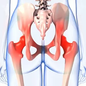
Background imageOrthopaedics Collection: Hip pain, artwork C013 / 5918

Hip pain, artwork C013 / 5918
Hip pain, computer artwork

Background imageOrthopaedics Collection: Knee realignment surgery, X-ray C016 / 6603

Knee realignment surgery, X-ray C016 / 6603
Knee alignment surgery. Coloured X-ray of the knee of a 55 year old overweightpatient that has undergone a wedge osteotomy to correct the alignment of the knee joint

Background imageOrthopaedics Collection: Knee realignment surgery, X-ray C016 / 6602

Knee realignment surgery, X-ray C016 / 6602
Knee alignment surgery. X-ray of the knee of a 55 year old overweightpatient that has undergone a wedge osteotomy to correct the alignment of the knee joint

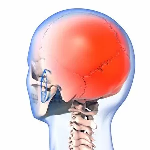
Background imageOrthopaedics Collection: Headache, artwork C013 / 5916

Headache, artwork C013 / 5916
Headache, computer artwork

Background imageOrthopaedics Collection: Headache, artwork C013 / 5915

Headache, artwork C013 / 5915
Headache, computer artwork

Background imageOrthopaedics Collection: Knee realignment surgery, X-ray C016 / 6599

Knee realignment surgery, X-ray C016 / 6599
Knee alignment surgery. Coloured X-ray of the knee of a 55 year old overweightpatient that has undergone a wedge osteotomy to correct the alignment of the knee joint

Background imageOrthopaedics Collection: Headache, artwork C013 / 5914

Headache, artwork C013 / 5914
Headache, computer artwork

Background imageOrthopaedics Collection: Headache, artwork C013 / 5913

Headache, artwork C013 / 5913
Headache, computer artwork

Background imageOrthopaedics Collection: Prosthetic knee and obesity, X-ray C016 / 6595

Prosthetic knee and obesity, X-ray C016 / 6595
Prosthetic knee and obesity. Coloured X-ray of a prosthetic knee joint (white) in an obese patient. This patient weighs 102 kilograms

Background imageOrthopaedics Collection: Elbow pain, artwork C013 / 5912

Elbow pain, artwork C013 / 5912
Elbow pain, computer artwork

Background imageOrthopaedics Collection: Prosthetic knee and obesity, X-ray C016 / 6594

Prosthetic knee and obesity, X-ray C016 / 6594
Prosthetic knee and obesity. Coloured X-ray of a prosthetic knee joint (white) in an obese patient. This patient weighs 102 kilograms

Background imageOrthopaedics Collection: Prosthetic knee and obesity, X-ray C016 / 6593

Prosthetic knee and obesity, X-ray C016 / 6593
Prosthetic knee and obesity. X-ray of a prosthetic knee joint (white) in an obese patient. This patient weighs 102 kilograms

Background imageOrthopaedics Collection: Ankle pain, artwork C013 / 5910

Ankle pain, artwork C013 / 5910
Ankle pain, computer artwork

Background imageOrthopaedics Collection: Pinned leg fracture, X-ray C016 / 6586

Pinned leg fracture, X-ray C016 / 6586
Pinned leg fracture. X-ray of the leg of a 33 year old patient that has been externally fixed after a skiing accident. Both the tibia (shine bone, upper bone) and fibula (calf bone)

Background imageOrthopaedics Collection: Fractured pelvis, CT scan C016 / 6584

Fractured pelvis, CT scan C016 / 6584
Fractured pelvis. Coloured computed tomography (CT) scan of the pelvis of a 56 year old that was in a road traffic accident

Background imageOrthopaedics Collection: Pinned leg fracture, X-ray C016 / 6585

Pinned leg fracture, X-ray C016 / 6585
Pinned leg fracture. X-ray of the leg of a 33 year old patient that has been externally fixed after a skiing accident. Both the tibia (shine bone, upper bone) and fibula (calf bone)

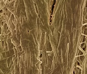
Background imageOrthopaedics Collection: Cortical bone, SEM C017 / 8496

Cortical bone, SEM C017 / 8496
False-coloured Scanning Electron Micrograph of cortical bone (compact bone). Cortical bone forms the dense, hard outer layer of most bones

Background imageOrthopaedics Collection: Salter Harris fracture, X-ray C017 / 8025

Salter Harris fracture, X-ray C017 / 8025
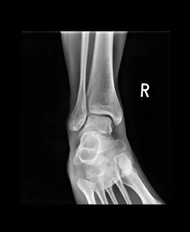
Salter Harris fracture. X-ray of the ankle of a child patient with a displaced Salter Harris fracture of the ankle. Salter Harris fractures occur at the epiphyseal, or growth

Background imageOrthopaedics Collection: Ankle fracture with bone cyst, X-ray C017 / 8016

Ankle fracture with bone cyst, X-ray C017 / 8016
Ankle fracture. X-ray of the fractured ankle of a patient. The bone had been weakened by a cyst, a fluid-filled cavity

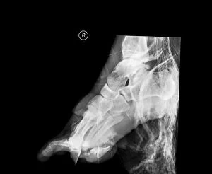
Background imageOrthopaedics Collection: Crushed right foot, X-ray C017 / 8012

Crushed right foot, X-ray C017 / 8012
Crushed right foot. X-ray of the right foot of an 18 year old male patient who has been in a motorcycle accident

Background imageOrthopaedics Collection: Amputated finger, X-ray C017 / 8010

Amputated finger, X-ray C017 / 8010
Amputated finger. X-ray of the hand of a 17 year old male patient who has had the tip of his middle finger amputated

Background imageOrthopaedics Collection: Broken wrist, X-ray C017 / 8008

Broken wrist, X-ray C017 / 8008
Broken wrist. X-ray of the arm of an 84 year old female patient with a fractured wrist

Background imageOrthopaedics Collection: Removed kneecap, X-ray C017 / 7981

Removed kneecap, X-ray C017 / 7981
Removed kneecap. X-ray of the knee of a 46 year old female that has had her kneecap (patella) surgically removed in a procedure known as a patellectomy

Background imageOrthopaedics Collection: Pinned jaw fracture, X-ray C017 / 7982

Pinned jaw fracture, X-ray C017 / 7982
Pinned jaw fracture. X-ray of the head of a 22 year old male patient who has had a fractured mandible (lower jaw bone) pinned (lower left)

Background imageOrthopaedics Collection: Broken elbow, X-ray C017 / 7979

Broken elbow, X-ray C017 / 7979
Broken elbow. X-ray of the elbow of a 4 year old female with a displaced supracondylar fracture. The fracture is at the base of the humerus, the upper arm bone (upper frame)

Background imageOrthopaedics Collection: Pinned broken ankle, X-ray C017 / 7978

Pinned broken ankle, X-ray C017 / 7978
Pinned broken ankle. X-ray of the ankle of a 32 year old male that has been pinned after a parachuting accident. The ankle was dislocated between the tibia (shin bone)

Background imageOrthopaedics Collection: Fractured and dislocated ankle, X-ray C017 / 7976

Fractured and dislocated ankle, X-ray C017 / 7976
Fractured and dislocated ankle. X-ray of the ankle of a 32 year old male who has been in a parachuting accident. The ankle is dislocated between the tibia (shin bone)

Background imageOrthopaedics Collection: Infected malunited fracture, X-ray C017 / 7972

Infected malunited fracture, X-ray C017 / 7972
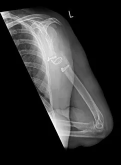
Infected malunited fracture. X-ray of the arm of a 65 year old female patient who has a bone infection after the nonunion of a fracture of the humerus (upper arm bone)

Background imageOrthopaedics Collection: Knuckle replacement, X-ray C017 / 7971

Knuckle replacement, X-ray C017 / 7971
Knuckle replacement. X-ray of the hand of a 41 year old male patient who has had two of the joints replaced between the hand bones (metacarpals) and the first finger bones (proximal phalanges)

Background imageOrthopaedics Collection: Broken elbow, X-ray C017 / 7968

Broken elbow, X-ray C017 / 7968
Broken elbow. X-ray of the elbow of a 5 year old female with a displaced supracondylar fracture. The fracture is at the base of the humerus, the upper arm bone (upper frame)

Background imageOrthopaedics Collection: Knuckle replacement, X-ray C017 / 7970

Knuckle replacement, X-ray C017 / 7970
Knuckle replacement. X-ray of the hand of a 41 year old male patient who has had two of the joints replaced between the hand bones (metacarpals) and the first finger bones (proximal phalanges)

Background imageOrthopaedics Collection: Pinned fractured elbow, X-ray C017 / 7969

Pinned fractured elbow, X-ray C017 / 7969
Pinned fractured elbow. X-ray of the elbow of a 5 year old female that has been internally fixed after a displaced supracondylar fracture

Background imageOrthopaedics Collection: Hospital hydrotherapy, 1920s C017 / 7871

Hospital hydrotherapy, 1920s C017 / 7871
Hospital hydrotherapy. Physiotherapy nurse (centre) supervising patients using water baths during a session of hydrotherapy, a form of physiotherapy (physical therapy)

Background imageOrthopaedics Collection: Joseph-Francois Malgaigne

Joseph-Francois Malgaigne (1806-1865) was a distinguished French surgeon, medical historian and anatomist. He specialised in fractures and the orthopaedic surgery of the knee, hip and shoulder

Background imageOrthopaedics Collection: Bone infection, X-ray C017 / 7861

Bone infection, X-ray C017 / 7861
Bone infection. X-ray of the foot of a diabetic patient osteomyelitis, a bacterial infection of the bone. Tissue damage to the foot caused by diabetes has lead to the infection

Background imageOrthopaedics Collection: Ankle fracture with bone cyst, X-ray C017 / 7862

Ankle fracture with bone cyst, X-ray C017 / 7862
Ankle fracture. X-ray of the fractured ankle of a patient. The bone had been weakened by a cyst, a fluid-filled cavity

Background imageOrthopaedics Collection: Ankle bone fracture, X-ray C017 / 7809

Ankle bone fracture, X-ray C017 / 7809
Ankle bone fracture. X-ray of the ankle of a patient with a fractured talus (ankle bone)

Background imageOrthopaedics Collection: Ankle bone fracture, X-ray C017 / 7808

Ankle bone fracture, X-ray C017 / 7808
Ankle bone fracture. X-ray of the ankle of a patient with a fractured talus (ankle bone, lower centre)

Background imageOrthopaedics Collection: Dislocated thumb, X-ray C017 / 7805

Dislocated thumb, X-ray C017 / 7805
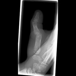
Dislocated thumb. X-ray of the right hand of a 10 year old male patient with a dislocated thumb

Background imageOrthopaedics Collection: Dislocated fingers, X-ray C017 / 7754

Dislocated fingers, X-ray C017 / 7754
Dislocated fingers. X-ray of the hand of a 60 year old male patient who has dislocated his third, fourth and fifth fingers

Background imageOrthopaedics Collection: Broken elbow, X-ray C017 / 7750

Broken elbow, X-ray C017 / 7750
Broken elbow. X-ray of the arm of a 44 year old female patient with a broken elbow

Background imageOrthopaedics Collection: Hospital hydrotherapy, 1920s C016 / 2383

Hospital hydrotherapy, 1920s C016 / 2383
Hospital hydrotherapy. Physiotherapy nurse (right) supervising a patient using a water bath during a session of hydrotherapy, a form of physiotherapy (physical therapy)

Background imageOrthopaedics Collection: Wrist pain, artwork C013 / 8822

Wrist pain, artwork C013 / 8822
Wrist pain, computer artwork

Background imageOrthopaedics Collection: Neck pain, artwork C013 / 8811

Neck pain, artwork C013 / 8811
Neck pain, computer artwork

Background imageOrthopaedics Collection: Lower back pain, artwork C013 / 8810

Lower back pain, artwork C013 / 8810
Lower back pain, computer artwork


All Professionally Made to Order for Quick Shipping

"Exploring the World of Orthopaedics: From Broken Bones to Artistic Marvels" A glimpse into orthopaedic challenges: X-ray reveals a broken wrist bone, reminding us of the intricate nature of healing. (X-ray C017 / 7187) The intersection of art and medicine: An exquisite artwork depicts the beauty of a hip replacement procedure, showcasing human resilience and innovation. Unveiling the hidden damage: Through an artistic representation, we witness the complexity of a damaged knee ligament and appreciate the expertise required for its restoration. Beyond physical pain: An intriguing X-ray artwork captures a headache, reminding us that orthopaedics encompasses not only bones but also various conditions affecting our well-being. Celebrating transformative surgeries: Another captivating artwork showcases a hip replacement, symbolizing hope and renewed mobility for countless individuals worldwide. Remembering Hugh Owen Thomas's legacy: Welsh surgeon C016 / 6299 revolutionized orthopaedics with his pioneering techniques, leaving an indelible mark on medical history. Peering beneath the surface: A jaw-dropping X-ray exposes a fractured jawbone, emphasizing both the fragility and strength inherent in our skeletal system. Historical insights into orthopaedic practices: An engraving from Traite des bandages et appareils illustrates a corset designed to prevent onanism—a testament to evolving medical beliefs in Paris during 1815 (b/w photo). Nurturing young lives with care: Dorothea Lange's poignant photograph captures a baby wearing homemade splints for club feet at an FSA camp in Tulare County, California in 1939—an early example of personalized orthopaedic treatment. Embracing knowledge through literature: The title page from L'Orthopedie ou l'Art de Prevenir et de Corriger dans les invites us to explore this comprehensive guide to preventing and correcting orthopaedic conditions.