Fraud Blocker Skip to main content

Orthopaedics Collection (page 3)

"Exploring the World of Orthopaedics: From Broken Bones to Artistic Marvels" A glimpse into orthopaedic challenges: X-ray reveals a broken wrist bone

Background imageOrthopaedics Collection: Prosthetic hip joint and Gruen zones C016 / 6779

Prosthetic hip joint and Gruen zones C016 / 6779
Prosthetic hip joint and Gruen zones. Cutaway diagram of a femur (thigh bone) showing a femoral component of a hip prosthesis and labels indicating Gruen zones

Background imageOrthopaedics Collection: Total hip replacement, X-rays

Total hip replacement, X-rays
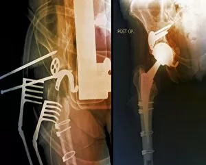
Total hip replacement. Coloured X-rays taken during (left) and after (right) surgery to replace the right hip joint of a 65-year-old patient using a prosthetic implant

Background imageOrthopaedics Collection: Failed hip replacement, CT scan

Failed hip replacement, CT scan
Failed hip replacement. Coloured computed tomography (CT) scan of a total hip replacement (solid white) in a 70-year-old patient

Background imageOrthopaedics Collection: Hip fracture reduction, 3D CT scan

Hip fracture reduction, 3D CT scan
Hip fracture reduction. Coloured 3D computed tomography (CT) scan of the pelvis of an 81-year-old patient with a fracture to the neck of the right femur (thigh bone)

Background imageOrthopaedics Collection: Fractured skull, X-ray C018 / 0591

Fractured skull, X-ray C018 / 0591
Fractured skull, X-ray. The fracture is the grey line at right that splits into two near the temple

Background imageOrthopaedics Collection: Dowagers hump, illustration

Dowagers hump, illustration
Dowagers hump. Illustration of a spine exhibiting hyperkyphosis, or dowagers hump. This is the concave curvature of the thoracic (chest) spine. It is most commonly caused by osteoporosis

Background imageOrthopaedics Collection: Broken elbow, X-ray C017 / 7584

Broken elbow, X-ray C017 / 7584
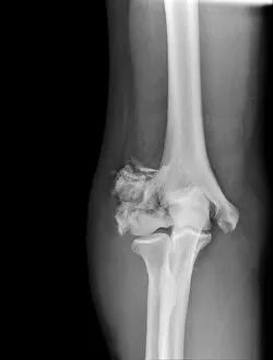
Broken elbow. X-ray of the arm of a 55 year old male patient with fractures to the elbow and radius (right-most lower arm bone)

Background imageOrthopaedics Collection: Broken arm, X-ray C017 / 7264

Broken arm, X-ray C017 / 7264
Broken arm. X-ray of the lower arm of a 6 year old male showing a greenstick fractures of the ulna (left) and radius (right) bones. Greenstick fractures occur when the bone bends and then cracks

Background imageOrthopaedics Collection: Broken jaw, X-ray C017 / 7558

Broken jaw, X-ray C017 / 7558
Broken jaw. X-ray of the head of a 22 year old male patient with a fractured mandible (lower jaw bone). The fracture is seen at left

Background imageOrthopaedics Collection: Louis Stromeyer, German surgeon, artwork C018 / 7052

Louis Stromeyer, German surgeon, artwork C018 / 7052
Louis Stromeyer (1804 - 1876). 1880 engraving of the German surgeon and physician Georg Friedrich Louis Stromeyer. Stromeyer was a pioneer in orthopaedics and orthopaedic surgery

Background imageOrthopaedics Collection: Scoliosis treatment, X-ray C017 / 7156

Scoliosis treatment, X-ray C017 / 7156
Scoliosis treatment. X-ray of the spine of a 46 year old female patient that has been fixed to treat scoliosis (sideways curvature)

Background imageOrthopaedics Collection: Radiologist examining a hand X-ray C014 / 1289

Radiologist examining a hand X-ray C014 / 1289
MODEL RELEASED. Radiologist examining a hand X-ray

Background imageOrthopaedics Collection: Facial fractures, X-ray C017 / 7638

Facial fractures, X-ray C017 / 7638
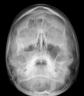
Facial fractures. X-ray of the skull of a 25 year old male patient with fractures to the right side (seen here on the left) of his face

Background imageOrthopaedics Collection: Fractured vertebra, X-ray C017 / 7585

Fractured vertebra, X-ray C017 / 7585
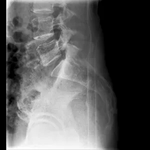
Fractured vertebra. X-ray of the lower spine of a 50 year old male with a fracture to the L1 lumbar vertebra (top centre)

Background imageOrthopaedics Collection: Fractured elbow, X-ray C017 / 7182

Fractured elbow, X-ray C017 / 7182
Fractured elbow. X-ray of the fractured left elbow of a 9 year old boy

Background imageOrthopaedics Collection: Dislocated wrist, X-ray C017 / 7563

Dislocated wrist, X-ray C017 / 7563
Dislocated wrist. X-ray of the wrist of a 16 year old male that has been dislocated

Background imageOrthopaedics Collection: Malunited fracture, X-ray C017 / 7641

Malunited fracture, X-ray C017 / 7641
Malunited fracture. X-ray of the leg of a 64 year old female patient with a malunited fracture of the femur (thigh bone). This occurs where a fracture fails to heal in the correct position

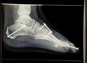
Background imageOrthopaedics Collection: Pinned foot bones after surgery, X-ray

Pinned foot bones after surgery, X-ray
Pinned foot bones after surgery. Lateral (side view) X-ray showing pins (white) inserted into the bones of a patients foot

Background imageOrthopaedics Collection: Hallux rigidus before surgery, X-ray

Hallux rigidus before surgery, X-ray
Hallux rigidus before surgery. X-ray showing foot bones before surgery to correct a case of hallux rigidus, a stiff big toe

Background imageOrthopaedics Collection: Fractured hip, artwork

Fractured hip, artwork
Fractured hip. Computer artwork of a fracture in the head of the femur (thigh bone)

Background imageOrthopaedics Collection: Paprosky femur defect, type IIIA lateral

Paprosky femur defect, type IIIA lateral
Paprosky femur defect. Cutaway artwork of bone degradation in a type IV medial-lateral femur cortex defect (Paprosky classification system)

Background imageOrthopaedics Collection: Paprosky femur defect, type IIIA med-lat

Paprosky femur defect, type IIIA med-lat
Paprosky femur defect. Cutaway artwork of bone degradation in a type IV lateral femur cortex defect (Paprosky classification system)

Background imageOrthopaedics Collection: Toothache, artwork C013 / 5927

Toothache, artwork C013 / 5927
Toothache, computer artwork

Background imageOrthopaedics Collection: Shoulder pain, artwork C013 / 5925

Shoulder pain, artwork C013 / 5925
Shoulder pain, computer artwork

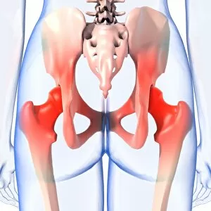
Background imageOrthopaedics Collection: Hip pain, artwork C013 / 5924

Hip pain, artwork C013 / 5924
Hip pain, computer artwork

Background imageOrthopaedics Collection: Toothache, artwork C013 / 5926

Toothache, artwork C013 / 5926
Toothache, computer artwork

Background imageOrthopaedics Collection: Lower back pain, artwork C013 / 5922

Lower back pain, artwork C013 / 5922
Lower back pain, computer artwork

Background imageOrthopaedics Collection: Neck pain, artwork C013 / 5923

Neck pain, artwork C013 / 5923
Neck pain, computer artwork

Background imageOrthopaedics Collection: Fractured and dislocated shoulder, X-ray C016 / 6607

Fractured and dislocated shoulder, X-ray C016 / 6607
Fractured and dislocated shoulder. Coloured X-ray of the shoulder of a 67 year old patient who has fallen and fractured the neck of the humerus (upper arm bone) and dislocated the shoulder

Background imageOrthopaedics Collection: Knee pain, artwork C013 / 5921

Knee pain, artwork C013 / 5921
Knee pain, computer artwork

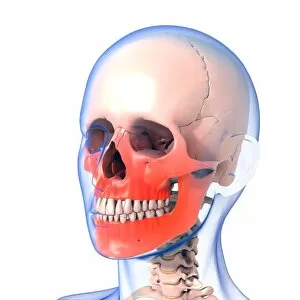
Background imageOrthopaedics Collection: Jaw pain, artwork C013 / 5920

Jaw pain, artwork C013 / 5920
Jaw pain, computer artwork

Background imageOrthopaedics Collection: Fractured and dislocated shoulder, X-ray C016 / 6606

Fractured and dislocated shoulder, X-ray C016 / 6606
Fractured and dislocated shoulder. Coloured X-ray of the shoulder of a 67 year old patient who has fallen and fractured the neck of the humerus (upper arm bone) and dislocated the shoulder

Background imageOrthopaedics Collection: Knee realignment surgery, X-ray C016 / 6604

Knee realignment surgery, X-ray C016 / 6604
Knee alignment surgery. Coloured X-ray of the knee of a 55 year old overweightpatient that has undergone a wedge osteotomy to correct the alignment of the knee joint

Background imageOrthopaedics Collection: Knee realignment surgery, X-ray C016 / 6601

Knee realignment surgery, X-ray C016 / 6601
Knee alignment surgery. Coloured X-ray of the knee of a 55 year old overweightpatient that has undergone a wedge osteotomy to correct the alignment of the knee joint

Background imageOrthopaedics Collection: Hip pain, artwork C013 / 5918

Hip pain, artwork C013 / 5918
Hip pain, computer artwork

Background imageOrthopaedics Collection: Knee realignment surgery, X-ray C016 / 6603

Knee realignment surgery, X-ray C016 / 6603
Knee alignment surgery. Coloured X-ray of the knee of a 55 year old overweightpatient that has undergone a wedge osteotomy to correct the alignment of the knee joint

Background imageOrthopaedics Collection: Knee realignment surgery, X-ray C016 / 6602

Knee realignment surgery, X-ray C016 / 6602
Knee alignment surgery. X-ray of the knee of a 55 year old overweightpatient that has undergone a wedge osteotomy to correct the alignment of the knee joint

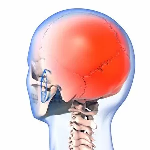
Background imageOrthopaedics Collection: Headache, artwork C013 / 5916

Headache, artwork C013 / 5916
Headache, computer artwork

Background imageOrthopaedics Collection: Headache, artwork C013 / 5915

Headache, artwork C013 / 5915
Headache, computer artwork

Background imageOrthopaedics Collection: Knee realignment surgery, X-ray C016 / 6599

Knee realignment surgery, X-ray C016 / 6599
Knee alignment surgery. Coloured X-ray of the knee of a 55 year old overweightpatient that has undergone a wedge osteotomy to correct the alignment of the knee joint

Background imageOrthopaedics Collection: Headache, artwork C013 / 5914

Headache, artwork C013 / 5914
Headache, computer artwork

Background imageOrthopaedics Collection: Headache, artwork C013 / 5913

Headache, artwork C013 / 5913
Headache, computer artwork

Background imageOrthopaedics Collection: Prosthetic knee and obesity, X-ray C016 / 6595

Prosthetic knee and obesity, X-ray C016 / 6595
Prosthetic knee and obesity. Coloured X-ray of a prosthetic knee joint (white) in an obese patient. This patient weighs 102 kilograms

Background imageOrthopaedics Collection: Elbow pain, artwork C013 / 5912

Elbow pain, artwork C013 / 5912
Elbow pain, computer artwork

Background imageOrthopaedics Collection: Prosthetic knee and obesity, X-ray C016 / 6594

Prosthetic knee and obesity, X-ray C016 / 6594
Prosthetic knee and obesity. Coloured X-ray of a prosthetic knee joint (white) in an obese patient. This patient weighs 102 kilograms

Background imageOrthopaedics Collection: Prosthetic knee and obesity, X-ray C016 / 6593

Prosthetic knee and obesity, X-ray C016 / 6593
Prosthetic knee and obesity. X-ray of a prosthetic knee joint (white) in an obese patient. This patient weighs 102 kilograms

Background imageOrthopaedics Collection: Ankle pain, artwork C013 / 5910

Ankle pain, artwork C013 / 5910
Ankle pain, computer artwork

Background imageOrthopaedics Collection: Pinned leg fracture, X-ray C016 / 6586

Pinned leg fracture, X-ray C016 / 6586
Pinned leg fracture. X-ray of the leg of a 33 year old patient that has been externally fixed after a skiing accident. Both the tibia (shine bone, upper bone) and fibula (calf bone)


All Professionally Made to Order for Quick Shipping

"Exploring the World of Orthopaedics: From Broken Bones to Artistic Marvels" A glimpse into orthopaedic challenges: X-ray reveals a broken wrist bone, reminding us of the intricate nature of healing. (X-ray C017 / 7187) The intersection of art and medicine: An exquisite artwork depicts the beauty of a hip replacement procedure, showcasing human resilience and innovation. Unveiling the hidden damage: Through an artistic representation, we witness the complexity of a damaged knee ligament and appreciate the expertise required for its restoration. Beyond physical pain: An intriguing X-ray artwork captures a headache, reminding us that orthopaedics encompasses not only bones but also various conditions affecting our well-being. Celebrating transformative surgeries: Another captivating artwork showcases a hip replacement, symbolizing hope and renewed mobility for countless individuals worldwide. Remembering Hugh Owen Thomas's legacy: Welsh surgeon C016 / 6299 revolutionized orthopaedics with his pioneering techniques, leaving an indelible mark on medical history. Peering beneath the surface: A jaw-dropping X-ray exposes a fractured jawbone, emphasizing both the fragility and strength inherent in our skeletal system. Historical insights into orthopaedic practices: An engraving from Traite des bandages et appareils illustrates a corset designed to prevent onanism—a testament to evolving medical beliefs in Paris during 1815 (b/w photo). Nurturing young lives with care: Dorothea Lange's poignant photograph captures a baby wearing homemade splints for club feet at an FSA camp in Tulare County, California in 1939—an early example of personalized orthopaedic treatment. Embracing knowledge through literature: The title page from L'Orthopedie ou l'Art de Prevenir et de Corriger dans les invites us to explore this comprehensive guide to preventing and correcting orthopaedic conditions.