Fraud Blocker Skip to main content

Nucleus Collection (page 10)

"The Nucleus: Unveiling the Mysteries of Life's Command Center" Exploring the intricate world within our cerebellum tissue

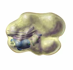
Background imageNucleus Collection: Golgi apparatus, artwork F006 / 9189

Golgi apparatus, artwork F006 / 9189
Computer artwork of the Golgi apparatus of the human cell. This organelle functions as a central delivery system for the cell

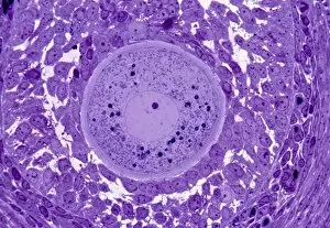
Background imageNucleus Collection: Oocyte, light micrograph

Oocyte, light micrograph
Oocyte. Light micrograph of a section through an oocyte within a early antral follicle in an ovary. Oocytes are immature ova, or egg cells

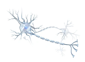
Background imageNucleus Collection: Nerve cells, artwork C015 / 4265

Nerve cells, artwork C015 / 4265
Nerve cells. Computer artwork of two nerve cells or neurons. Neurons are responsible for passing information around the central nervous system (CNS) and from the CNS to the rest of the body

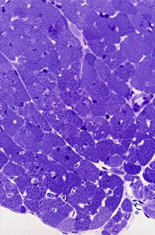
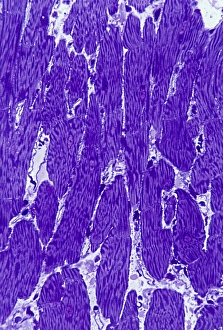
Background imageNucleus Collection: Cardiac muscle, light micrograph

Cardiac muscle, light micrograph
Cardiac muscle. Light micrograph of a section through muscle tissue from a heart, showing the cardiomyocyte cells. Heart muscle cells show central nuclei but the dominant feature is the abundance of

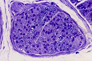
Background imageNucleus Collection: Peripheral nerve, light micrograph

Peripheral nerve, light micrograph
Peripheral nerve. Light micrograph of a section through a peripheral nerve. This is a mixed nerve with myelinated axons (dark blue circles)

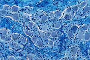
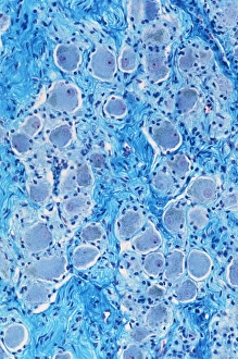
Background imageNucleus Collection: Nerve ganglion, light micrograph

Nerve ganglion, light micrograph
Nerve ganglion. Light micrograph of a section through a dorsal (sensory) spinal root ganglion associated with a sensory nerve root of the spinal cord. Sensory information from peripheral sites e.g

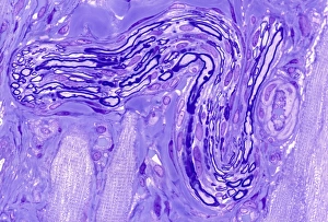
Background imageNucleus Collection: Myelinated nerve, light micrograph

Myelinated nerve, light micrograph
Myelinated nerve. Light micrograph of a section through a peripheral myelinated nerve, showing many individual axons each covered with deep blue-staining myelin sheaths

Background imageNucleus Collection: Fertilisation, artwork C016 / 7516

Fertilisation, artwork C016 / 7516
Fertilisation. Cutaway computer artwork of a sperm cell (spermatozoa) penetrating an egg (large). The nucleus of the sperm cell will fuse with the egg and its DNA (deoxyribonucleic acid)

Background imageNucleus Collection: DNA packaging, artwork C016 / 7517

DNA packaging, artwork C016 / 7517
DNA packaging. Computer artwork showing how DNA (deoxyribonucleic acid) is packaged within cells. Two DNA strands, consisting of a sugar-phosphate backbone attached to nucleotide bases

Background imageNucleus Collection: Corpus luteum, TEM

Corpus luteum, TEM
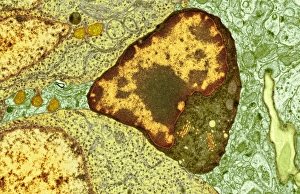
Corpus luteum. Transmission electron micrograph (TEM) of a section through the corpus luteum of an ovary, showing several luteal cells

Background imageNucleus Collection: Comet over trees, artwork C015 / 0777

Comet over trees, artwork C015 / 0777
Comet over trees, artwork. Comets are bodies of ice and dust that enter the inner solar system from the outer solar system

Background imageNucleus Collection: Ductal breast cancer, TEM

Ductal breast cancer, TEM
Ductal breast cancer. Transmission electron micrograph (TEM) of a section through breast tissue, showing malignant (cancerous) epithelial cells arising from the milk ducts of the breast

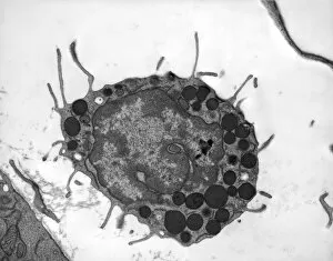
Background imageNucleus Collection: Macrophage white blood cells, TEM

Macrophage white blood cells, TEM
Macrophage white blood cells. Transmission electron micrograph (TEM) of a section through two macrophages located within connective tissue, showing the nuclei and numerous lysosomes (dark spots)

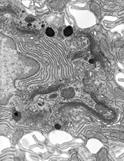
Background imageNucleus Collection: Pancreatic acinar cell, TEM

Pancreatic acinar cell, TEM
Pancreatic acinar cell. Transmission electron micrograph (TEM) of a section through an enzyme-secreting acinar cell in the human pancreas, showing the nucleus (dark purple, centre)

Background imageNucleus Collection: Basophil white blood cell, TEM

Basophil white blood cell, TEM
Basophil white blood cell. Transmission electron micrograph (TEM) of section through a circulating basophil cell. Basophils are the smallest and least common of the white blood cells

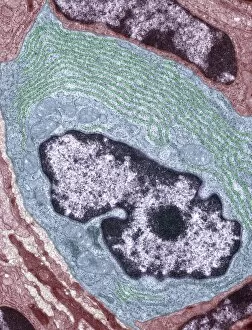
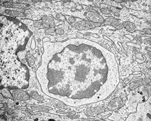
Background imageNucleus Collection: Lymphocyte white blood cell, TEM

Lymphocyte white blood cell, TEM
Lymphocyte white blood cell. Transmission electron micrograph (TEM) of a section through lymphocyte, showing the nucleus (large, round, centre), endoplasmic reticulum (ER)

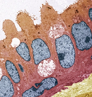
Background imageNucleus Collection: Enterocyte, TEM

Enterocyte, TEM
Enterocyte. Transmission electron micrograph (TEM) of a section through the cytoplasm and part of the nucleus of an enterocyte cell located in a crypt of Lieberkuhn of the small intestine

Background imageNucleus Collection: Comet and Sun, artwork C015 / 0776

Comet and Sun, artwork C015 / 0776
Comet and Sun, artwork. Comets are bodies of ice and dust that enter the inner solar system from the outer solar system. As they approach the Sun, the heat boils the ice

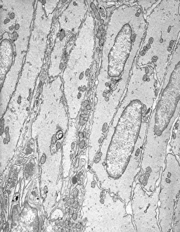
Background imageNucleus Collection: Smooth muscle tissue, TEM

Smooth muscle tissue, TEM
Smooth muscle tissue. Transmission electron micrograph (TEM) of a longitudinal section through smooth muscle cells. Smooth muscle cells are spindle-shaped and often closely packed together

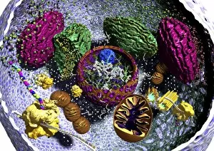
Background imageNucleus Collection: Eukaryote cell, artwork C016 / 6260

Eukaryote cell, artwork C016 / 6260
Eukaryote cell. Computer artwork showing the internal structure of a typical eukaryotic cell. Eukaryotes are organisms whose cells contain a membrane-bound nucleus (karyon, orange)

Background imageNucleus Collection: Prokaryote and eukaryote cells, artwork C016 / 6259

Prokaryote and eukaryote cells, artwork C016 / 6259
Prokaryote and eukaryote cells. Computer artwork comparing typical prokaryotic (top) and eukaryotic (bottom) cells. Prokaryotes are organisms whose cells lack a membrane-bound nucleus (karyon)

Background imageNucleus Collection: Heart muscle, light micrograph C016 / 0517

Heart muscle, light micrograph C016 / 0517
Heart muscle. Light micrograph of a section through heart (cardiac) muscle tissue, showing cardiomyocyte muscle cells. These cells have a centrally located nucleus and are branched

Background imageNucleus Collection: Nerve ganglion, light micrograph C016 / 0532

Nerve ganglion, light micrograph C016 / 0532
Nerve ganglion. Light micrograph of a section through a nerve ganglion of the peripheral nervous system, showing clusters of nerve cell bodies

Background imageNucleus Collection: Myelinated nerve fibres, light micrograph C016 / 0531

Myelinated nerve fibres, light micrograph C016 / 0531
Myelinated nerve fibres. Light micrograph of a section through a spinal sensory nerve containing many closely packed axons exhibiting a wave-type appearance

Background imageNucleus Collection: Neutrophil white blood cell, illustration C018 / 0741

Neutrophil white blood cell, illustration C018 / 0741
Neutrophil white blood cell, illustration. Neutrophils are the most abundant white blood cell in humans. They have a multi-lobed nucleus (dark purple)

Background imageNucleus Collection: Animal cell, illustration C018 / 0734

Animal cell, illustration C018 / 0734
Animal cell. Illustration showing the organelles present in an animal cell. At lower centre is the nucleus, which contains the cells genetic information in the form of DNA (deoxyribonucleic acid)

Background imageNucleus Collection: DNA packaging, illustration C018 / 0747

DNA packaging, illustration C018 / 0747
DNA packaging. Illustration showing how DNA (deoxyribonucleic acid) is packaged within cells. Two DNA strands, consisting of a sugar-phosphate backbone attached to nucleotide bases

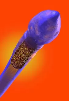
Background imageNucleus Collection: Sperm cell, artwork C018 / 6996

Sperm cell, artwork C018 / 6996
Sperm cell anatomy. Cutaway computer artwork showing the internal structure of a sperm cell (spermatozoon), the male sex cell

Background imageNucleus Collection: Intestinal gland cell, TEM C014 / 1442

Intestinal gland cell, TEM C014 / 1442
Intestinal gland cell. Transmission electron micrograph (TEM) of a section through an undifferentiated cell from the intestinal tract, showing extensive rough endoplasmic reticulum (ER)

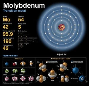
Background imageNucleus Collection: Rutherfordium, atomic structure

Rutherfordium, atomic structure
Argon (Ar). Diagram of the nuclear composition, electron configuration, chemical data, and valence orbitals of an atom of argon-40 (atomic number: 18)

Background imageNucleus Collection: Monocyte white blood cell, TEM

Monocyte white blood cell, TEM
Monocyte white blood cell. Transmission electron micrograph (TEM) of a section through a monocyte white blood cell, showing the cell nucleus (blue)

Background imageNucleus Collection: Animal cell organelles, artwork

Animal cell organelles, artwork
Animal cell organelles. Artwork showing the organelles in a eukaryotic cell. This is an animal cell. Structures include the nucleus (centre) which has a membrane with nuclear pores (purple)

Background imageNucleus Collection: Fat cell anatomy, artwork

Fat cell anatomy, artwork
Fat cell anatomy. Artwork of an adipocyte (fat cell) and its internal organelles. The majority of the cell is filled with a lipid (fat) droplet

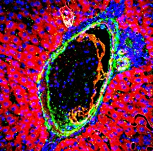
Background imageNucleus Collection: Liver portal triad, light micrograph C016 / 8490

Liver portal triad, light micrograph C016 / 8490
Liver portal triad. Fluorescence deconvolution micrograph of a section through a portal triad in liver tissue, showing hepatocyte cells (red)

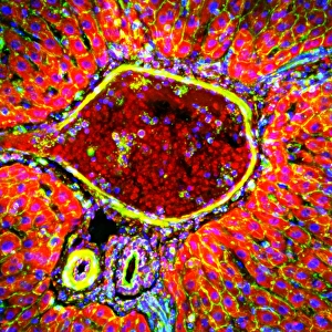
Background imageNucleus Collection: Liver portal triad, light micrograph C016 / 8489

Liver portal triad, light micrograph C016 / 8489
Liver portal triad. Fluorescence deconvolution micrograph of a section through a portal triad in liver tissue, showing hepatocyte cells (red)

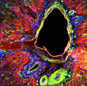
Background imageNucleus Collection: Liver portal triad, light micrograph C016 / 8488

Liver portal triad, light micrograph C016 / 8488
Liver portal triad. Fluorescence deconvolution micrograph of a section through a portal triad in liver tissue, showing hepatocyte cells (red)

Background imageNucleus Collection: Kidney tissue, fluorescence micrograph C016 / 8484

Kidney tissue, fluorescence micrograph C016 / 8484
Kidney tissue. Fluorescence deconvolution micrograph of a section through a kidney, showing glomeruli (green), cell nuclei (blue dots), and renal tubules (red, circular)

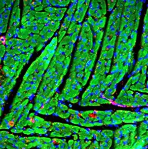
Background imageNucleus Collection: Heart muscle, fluorescence micrograph C016 / 8483

Heart muscle, fluorescence micrograph C016 / 8483
Heart muscle. Fluorescence deconvolution micrograph of a section through heart tissue, showing the angular distribution of the myocardium (cardiac muscle) fibres (green)

Background imageNucleus Collection: Heart muscle, fluorescence micrograph C016 / 8481

Heart muscle, fluorescence micrograph C016 / 8481
Heart muscle. Fluorescence deconvolution micrograph of a section through heart tissue, showing blood vessels (oval, centre-left and upper centre) running through the myocardium (cardiac muscle)

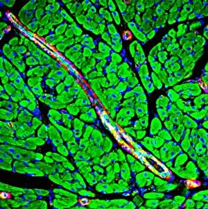
Background imageNucleus Collection: Heart muscle, fluorescence micrograph C016 / 8478

Heart muscle, fluorescence micrograph C016 / 8478
Heart muscle. Fluorescence deconvolution micrograph of a section through heart tissue, showing a blood vessel (diagonal, centre) running through the myocardium (cardiac muscle, green)

Background imageNucleus Collection: Heart muscle, fluorescence micrograph C016 / 8477

Heart muscle, fluorescence micrograph C016 / 8477
Heart muscle. Fluorescence deconvolution micrograph of a section through heart tissue, showing a blood vessel (blue, diagonal, centre) running through the myocardium (cardiac muscle, green)

Background imageNucleus Collection: HIV invading white blood cell, artwork

HIV invading white blood cell, artwork
HIV invading white blood cell. Computer artwork showing HIV (human immunodeficiency virus) particles (virions, purple) invading a white blood cell (large, centre) in the human bloodstream

Background imageNucleus Collection: Hydrogen atom, conceptual model C013 / 5605

Hydrogen atom, conceptual model C013 / 5605
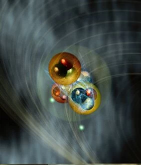
Hydrogen atom, conceptual model. Computer artwork representing the atomic structure of hydrogen. Hydrogen has one proton and one neutron (large spheres) in its nucleus (large circle, centre)

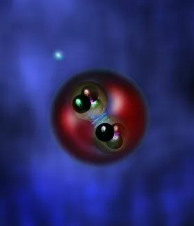
Background imageNucleus Collection: Helium atom, conceptual model C013 / 5600

Helium atom, conceptual model C013 / 5600
Helium atom, conceptual model. Computer artwork representing the atomic structure of helium. Helium has two protons and two neutrons (large spheres) in its nucleus (faint circle, centre)

Background imageNucleus Collection: Helium atom, conceptual model C013 / 5601

Helium atom, conceptual model C013 / 5601
Helium atom, conceptual model. Computer artwork representing the atomic structure of helium. Helium has two protons and two neutrons (large spheres) in its nucleus (faint circle, centre)

Background imageNucleus Collection: Brain nerve cells, TEM C014 / 0356

Brain nerve cells, TEM C014 / 0356
Brain nerve cells. Transmission electron micrograph (TEM) of a section through brain tissue from the cerebral cortex, showing numerous neurons (nerve cells) surrounded by axons and dendrites

Background imageNucleus Collection: Brain cell, TEM C014 / 0358

Brain cell, TEM C014 / 0358
Brain cell. Transmission electron micrograph (TEM) of a section through an oligodendrocyte in human brain tissue. Oligodendrocytes occur in both the white

Background imageNucleus Collection: Brain cell, TEM C014 / 0359

Brain cell, TEM C014 / 0359
Brain cell. Transmission electron micrograph (TEM) of a section through an oligodendrocyte in human brain tissue. Oligodendrocytes occur in both the white


All Professionally Made to Order for Quick Shipping

"The Nucleus: Unveiling the Mysteries of Life's Command Center" Exploring the intricate world within our cerebellum tissue, where the nucleus orchestrates every movement and balance. A mesmerizing light micrograph reveals the nucleus at the heart of M51 Whirlpool Galaxy, a cosmic dance guided by its gravitational pull. Witnessing life's beginning through a budding yeast cell, as its nucleus holds the blueprint for growth and reproduction. Delving into the secrets of communication with a nerve cell captured in stunning detail through SEM imaging, showcasing its complex nucleus. Behold the majestic Sombrero Galaxy (Messier 104), where billions of stars revolve around their central nucleus like celestial dancers in perfect harmony. The hippocampus brain tissue unveils memories etched within each neuron's nucleus, shaping who we are and what we remember. Peering into HeLa cells under a light microscope (C017/8299), revealing their remarkable nuclei that have revolutionized medical research. An artistic representation takes us deep into Medulla Oblongata's core—the vital center controlling essential bodily functions—where nuclei reign supreme. Captivating artwork depicts nuclear fission—a powerful force harnessed to generate energy while reminding us of its immense potential and responsibility. Marvel at NGC 4258 spiral galaxy's galactic light show, where vibrant colors illuminate star clusters revolving around their radiant nuclear core. Human cells come alive under scrutiny as their nuclei hold genetic information that shapes our unique characteristics and existence itself. Exploring kidney tubules in section unravels an intricate network governed by countless tiny nuclei working harmoniously to maintain our body’s equilibrium. Intricate yet awe-inspiring, these glimpses into various realms remind us that no matter how vast or minuscule, from galaxies to microscopic cells - all bow to the commanding presence of the nucleus.