Fraud Blocker Skip to main content

Neurology Collection (page 9)

Neurology, the fascinating study of the nervous system, unravels the intricate workings of our brain and its various components

Background imageNeurology Collection: Anatomy of human skull, eyeball and arteries to brain

Anatomy of human skull, eyeball and arteries to brain

Background imageNeurology Collection: Albrecht von Haller (1708-1777), Swiss physician and scientist who was the founder of neurology

Albrecht von Haller (1708-1777), Swiss physician and scientist who was the founder of neurology, c1770. Experimental physiology. Engraving c1790

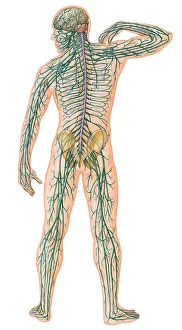
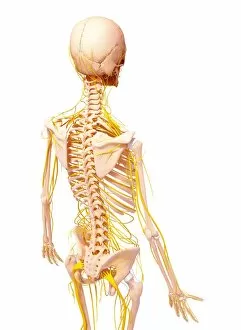
Background imageNeurology Collection: Rear view of Male Body with Major Nerves Superimposed

Rear view of Male Body with Major Nerves Superimposed

Background imageNeurology Collection: Human brain

Human brain

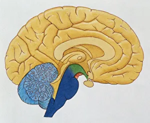
Background imageNeurology Collection: Basic cutaway illustration of the human brain showing the thalamus, cerebrum, hypothalmus

Basic cutaway illustration of the human brain showing the thalamus, cerebrum, hypothalmus, cerebellum and brain stem

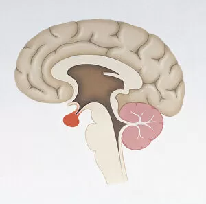
Background imageNeurology Collection: Illustrated diagram of the brain

Illustrated diagram of the brain

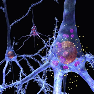
Background imageNeurology Collection: Prion disease treatment, artwork C018 / 1838

Prion disease treatment, artwork C018 / 1838
Prion disease treatment. Artwork of three pyramidal neurons (nerve cells). The cell at left is healthy. The cell at back has died because of a prion (pink) infection

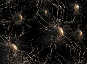
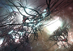
Background imageNeurology Collection: Neural network, artwork C017 / 2276

Neural network, artwork C017 / 2276
Neural network, computer artwork

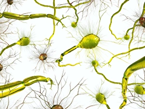
Background imageNeurology Collection: Astrocyte nerve cells, artwork C017 / 3425

Astrocyte nerve cells, artwork C017 / 3425
Astrocyte nerve cells, computer artwork. Astrocytes are a type of glial cell.They provide structural support and protection for neurons (nerve cells) and also supply them with nutrients and oxygen

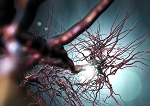
Background imageNeurology Collection: Pyramidal nerve cells, artwork C017 / 2273

Pyramidal nerve cells, artwork C017 / 2273
Pyramidal nerve cells. Computer artwork of pyramidal nerve cells from the cerebral cortex of the brain. Pyramidal cells are so named for their triangular cell bodies

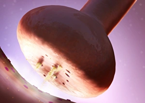
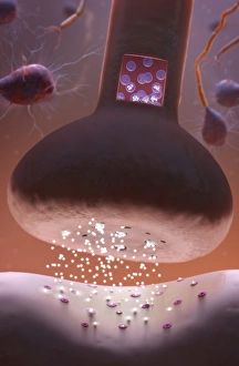
Background imageNeurology Collection: Nerve synapse, artwork C017 / 3427

Nerve synapse, artwork C017 / 3427
Nerve synapse. Computer artwork of of a junction, or synapse, between two nerve cells (neurons). As the electrical signal reaches the presynaptic end of a neuron it triggers the release of

Background imageNeurology Collection: Brain scan, conceptual image C017 / 7392

Brain scan, conceptual image C017 / 7392
MODEL RELEASED. Brain scan, conceptual composite image

Background imageNeurology Collection: Bennet percussor, circa 1860 C017 / 6934

Bennet percussor, circa 1860 C017 / 6934
Bennet percussor with ivory pleximeter dating from 1860s, England. A percussor is a small hammer used for testing reflexes and for percussive examinations of the chest

Background imageNeurology Collection: Brain scan, conceptual image C017 / 7394

Brain scan, conceptual image C017 / 7394
MODEL RELEASED. Brain scan, conceptual composite image

Background imageNeurology Collection: Brain scan, conceptual image C017 / 7398

Brain scan, conceptual image C017 / 7398
MODEL RELEASED. Brain scan, conceptual composite image

Background imageNeurology Collection: Brain scan, conceptual image C017 / 7397

Brain scan, conceptual image C017 / 7397
MODEL RELEASED. Brain scan, conceptual composite image

Background imageNeurology Collection: Bennet percussor, circa 1860 C017 / 6971

Bennet percussor, circa 1860 C017 / 6971
Bennet percussor dating from 1860s, England. A percussor is a small hammer used for testing reflexes and for percussive examinations of the chest

Background imageNeurology Collection: Nerve synapse, artwork C017 / 3428

Nerve synapse, artwork C017 / 3428
Nerve synapse. Computer artwork of of a junction, or synapse, between two nerve cells (neurons). As the electrical signal reaches the presynaptic end of a neuron it triggers the release of

Background imageNeurology Collection: Corpus callosum, DTI MRI scan C017 / 7044

Corpus callosum, DTI MRI scan C017 / 7044
Corpus callosum. Coronal 3D diffusion tensor imaging (DTI) magnetic resonance imaging (MRI) scan of nerve pathways (coloured) in and around the brains corpus callosum

Background imageNeurology Collection: SIRT3 molecule, artwork C017 / 3657

SIRT3 molecule, artwork C017 / 3657
SIRT3 molecule. Computer artwork showing the structure of a molecule of NAD-dependent deacetylase sirtuin-3, mitochondrial (SIRT3)

Background imageNeurology Collection: Pyramidal nerve cells, artwork C017 / 2274

Pyramidal nerve cells, artwork C017 / 2274
Pyramidal nerve cells. Computer artwork of pyramidal nerve cells from the cerebral cortex of the brain. Pyramidal cells are so named for their triangular cell bodies

Background imageNeurology Collection: Brain scan, conceptual image C017 / 7391

Brain scan, conceptual image C017 / 7391
MODEL RELEASED. Brain scan, conceptual composite image

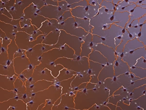
Background imageNeurology Collection: Neural network, artwork C017 / 2278

Neural network, artwork C017 / 2278
Neural network, computer artwork

Background imageNeurology Collection: Synaesthesia, conceptual artwork C017 / 7072

Synaesthesia, conceptual artwork C017 / 7072
Synaesthesia, conceptual artwork. Synaesthesia is a neurological condition where stimulation of one sensory or cognitive pathway leads to automatic

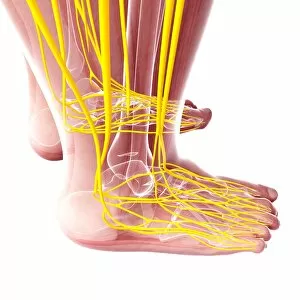
Background imageNeurology Collection: Human foot nervous system, artwork F007 / 1148

Human foot nervous system, artwork F007 / 1148
Human foot nervous system, computer artwork

Background imageNeurology Collection: Female nervous system, artwork F007 / 4284

Female nervous system, artwork F007 / 4284
Female nervous system, computer artwork

Background imageNeurology Collection: Female nervous system, artwork F007 / 3461

Female nervous system, artwork F007 / 3461
Female nervous system, computer artwork

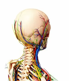
Background imageNeurology Collection: Human nervous system, artwork F007 / 3986

Human nervous system, artwork F007 / 3986
Human nervous system, computer artwork

Background imageNeurology Collection: Human nervous system, artwork F007 / 3394

Human nervous system, artwork F007 / 3394
Human nervous system, computer artwork

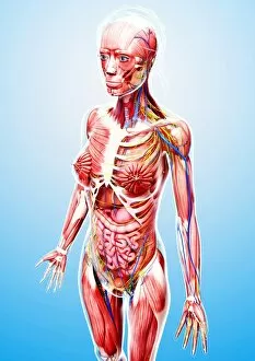
Background imageNeurology Collection: Female anatomy, artwork F007 / 3969

Female anatomy, artwork F007 / 3969
Female anatomy, computer artwork

Background imageNeurology Collection: Female anatomy, artwork F007 / 3665

Female anatomy, artwork F007 / 3665
Female anatomy, computer artwork

Background imageNeurology Collection: Human nervous system, artwork F007 / 4283

Human nervous system, artwork F007 / 4283
Human nervous system, computer artwork

Background imageNeurology Collection: Female nervous system, artwork F007 / 4298

Female nervous system, artwork F007 / 4298
Female nervous system, computer artwork

Background imageNeurology Collection: Human nervous system, artwork F007 / 4193

Human nervous system, artwork F007 / 4193
Human nervous system, computer artwork

Background imageNeurology Collection: Female anatomy, artwork F007 / 4084

Female anatomy, artwork F007 / 4084
Female anatomy, computer artwork

Background imageNeurology Collection: Female anatomy, artwork F007 / 3976

Female anatomy, artwork F007 / 3976
Female anatomy, computer artwork

Background imageNeurology Collection: Female nervous system, artwork F007 / 3589

Female nervous system, artwork F007 / 3589
Female nervous system, computer artwork

Background imageNeurology Collection: Human nervous system, artwork F007 / 4085

Human nervous system, artwork F007 / 4085
Human nervous system, computer artwork

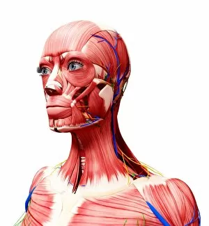
Background imageNeurology Collection: Human anatomy, artwork F007 / 3686

Human anatomy, artwork F007 / 3686
Human anatomy, computer artwork

Background imageNeurology Collection: Female anatomy, artwork F007 / 3768

Female anatomy, artwork F007 / 3768
Female anatomy, computer artwork

Background imageNeurology Collection: Female anatomy, artwork F007 / 3868

Female anatomy, artwork F007 / 3868
Female anatomy, computer artwork

Background imageNeurology Collection: Human anatomy, artwork F007 / 3471

Human anatomy, artwork F007 / 3471
Human anatomy, computer artwork

Background imageNeurology Collection: Female nervous system, artwork F007 / 3870

Female nervous system, artwork F007 / 3870
Female nervous system, computer artwork

Background imageNeurology Collection: Female nervous system, artwork F007 / 3574

Female nervous system, artwork F007 / 3574
Female nervous system, computer artwork

Background imageNeurology Collection: Human anatomy, artwork F007 / 3463

Human anatomy, artwork F007 / 3463
Human anatomy, computer artwork

Background imageNeurology Collection: Female anatomy, artwork F007 / 3771

Female anatomy, artwork F007 / 3771
Female anatomy, computer artwork

Background imageNeurology Collection: Female anatomy, artwork F007 / 3565

Female anatomy, artwork F007 / 3565
Female anatomy, computer artwork

Background imageNeurology Collection: Human anatomy, artwork F007 / 4090

Human anatomy, artwork F007 / 4090
Human anatomy, computer artwork


All Professionally Made to Order for Quick Shipping

Neurology, the fascinating study of the nervous system, unravels the intricate workings of our brain and its various components. From the Motor homunculus model that depicts how different body parts are represented in our motor cortex to the Histological Diagram of a Mammalian Retina revealing the complexity of our visual processing, every aspect is awe-inspiring. The Cerebellum tissue, as seen through a light micrograph, showcases its unique structure responsible for coordinating movement and balance. Meanwhile, an inferior view of the Anatomy of human brain highlights its remarkable organization and interconnectedness. Through Brain fibres captured by DTI MRI scans like C017 / 7099 or C017 / 7035, we gain insights into neural pathways that facilitate communication between different regions. These pathways form a complex network crucial for transmitting information throughout our brain. Exploring Brain blood vessels using a 3D angiogram (C007 / 1981) reveals their vital role in supplying oxygen and nutrients to sustain neuronal function. Additionally, understanding Motor and sensory homunculi helps us comprehend how specific areas within our brains control different bodily functions. Delving deeper into neurology brings us face-to-face with Medulla oblongata artwork depicting this critical region involved in regulating essential autonomic functions such as breathing and heart rate. Furthermore, observing Nerve and glial cells under a light microscope illustrates their diverse roles in supporting neurons' health and functionality. Finally, examining Synapse nerve junctions through TEM unveils these microscopic structures where electrical signals are transmitted from one neuron to another—a fundamental process underlying all brain activity. In essence, neurology takes us on an incredible journey deep within ourselves—unveiling mysteries at both macroscopic levels like anatomical structures or microscopically exploring cellular intricacies, and is through this exploration that we gain profound knowledge about how our brains work—the very essence of who we are as individuals.