Fraud Blocker Skip to main content

Neurology Collection (page 7)

Neurology, the fascinating study of the nervous system, unravels the intricate workings of our brain and its various components

Background imageNeurology Collection: Metropolis (1927) C016 / 4491

Metropolis (1927) C016 / 4491
Metropolis (1927). Poster for the the 1927 film Metropolis by Austrian film-maker Fritz Lang (1890-1976), showing the character Maria in Rotwangs transformation machine

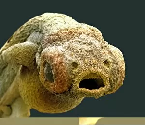
Background imageNeurology Collection: Zebra fish embryo, SEM C013 / 9587

Zebra fish embryo, SEM C013 / 9587
Zebra fish embryo. Coloured scanning electron micrograph (SEM) of the embryo of a zebrafish (Danio rerio), showing its eyes either side of its large open mouth (centre)

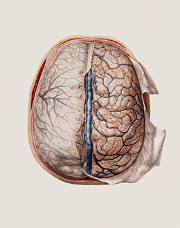
Background imageNeurology Collection: Brain meninges

Brain meninges, historical anatomical artwork. This cranial (top) view of the brain (with front at the top) and its surrounding protective meninges, which have been partially dissected away

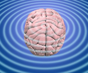
Background imageNeurology Collection: Brain waves, conceptual image

Brain waves, conceptual image
Brain waves. Conceptual image of waves emanating from a human brain. This can represent the electrical activity of the brain, as recorded on an electroencephalogram (EEG)
Background imageNeurology Collection: Foetal brain development, artwork

Foetal brain development, artwork. During the 4th week (upper left) the neural tube begins to differentiate into a spinal cord (green), forebrain (blue), midbrain (grey) and hindbrain (orange)

Background imageNeurology Collection: Spinal cord regions, diagram

Spinal cord regions, diagram
Spinal cord regions. Diagram of a sagittal section through the human skull and backbone, showing the five main regions of the spinal cord and the numbered nerves associated with them

Background imageNeurology Collection: Multiple sclerosis, MRI scan

Multiple sclerosis, MRI scan

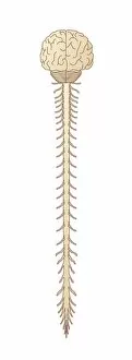
Background imageNeurology Collection: Brain and spinal cord, artwork

Brain and spinal cord, artwork
Brain and spinal cord. Computer artwork showing the brain (top) and segments of the spinal cord. The spinal cord begins at the medulla oblongata at the base of the brainstem

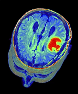
Background imageNeurology Collection: Brain tumour, 3D-MRI scan

Brain tumour, 3D-MRI scan
Brain tumour. Coloured three-dimensional model of a male head, made up from numerous magnetic resonance imaging (MRI) scans

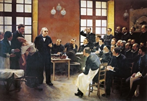
Background imageNeurology Collection: Charcot lectures at La Salpetriere by Pierre-Andre Brouillet (1857-1914)

Charcot lectures at La Salpetriere by Pierre-Andre Brouillet (1857-1914)
France - Lyon. HA

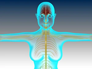
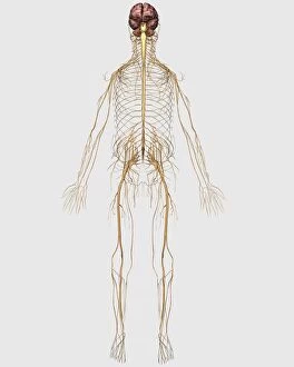
Background imageNeurology Collection: Conceptual image of female nervous system with brain

Conceptual image of female nervous system with brain

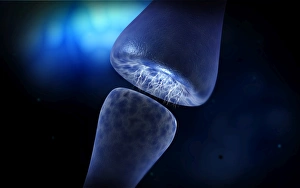
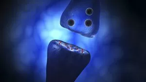
Background imageNeurology Collection: Conceptual image of synapse of neuron inside brain

Conceptual image of synapse of neuron inside brain

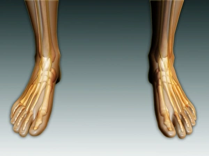
Background imageNeurology Collection: Conceptual image of human legs and feet with nervous system

Conceptual image of human legs and feet with nervous system

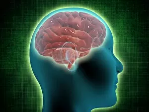
Background imageNeurology Collection: Conceptual image of female body with brain, side view

Conceptual image of female body with brain, side view

Background imageNeurology Collection: Main arteries and veins within a glass head

Main arteries and veins within a glass head

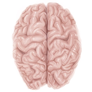
Background imageNeurology Collection: Superior view of human brain

Superior view of human brain

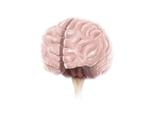
Background imageNeurology Collection: Brain surface anatomy

Brain surface anatomy

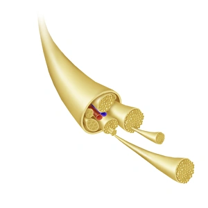
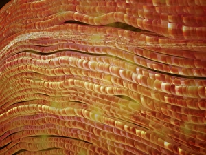
Background imageNeurology Collection: Detail of a nerve bundle

Detail of a nerve bundle

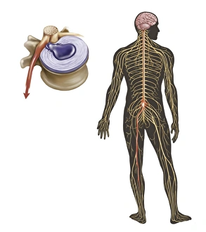
Background imageNeurology Collection: Sciatica caused from herniated disc

Sciatica caused from herniated disc

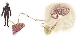
Background imageNeurology Collection: Pathway of a pain message via sensory nerve in injured muscle

Pathway of a pain message via sensory nerve in injured muscle, to pain gate in spinal cord to limbic system, frontal cortex and sensory cortex in the human brain

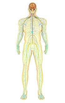
Background imageNeurology Collection: Anatomy of human nervous system and lymphatic system

Anatomy of human nervous system and lymphatic system, front view

Background imageNeurology Collection: Medical illustration of peripheral nervous system with brain

Medical illustration of peripheral nervous system with brain

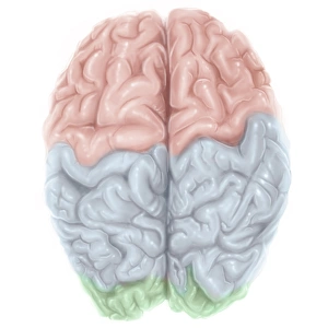
Background imageNeurology Collection: Superior view of human brain with colored lobes

Superior view of human brain with colored lobes

Background imageNeurology Collection: Anatomy of pituitary gland

Anatomy of pituitary gland

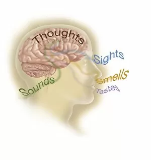
Background imageNeurology Collection: Representation of how our senses affect our thoughts

Representation of how our senses affect our thoughts

Background imageNeurology Collection: Conceptual image of synaptic vesicles

Conceptual image of synaptic vesicles

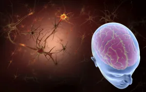
Background imageNeurology Collection: Conceptual image of human brain with neurons

Conceptual image of human brain with neurons in background

Background imageNeurology Collection: Anatomy of female body with arteries, veins and nervous system

Anatomy of female body with arteries, veins and nervous system

Background imageNeurology Collection: Male human head with skull and artificial electronic circuit brain

Male human head with skull and artificial electronic circuit brain in ghost effect, side view

Background imageNeurology Collection: Representation of how our thoughts affect our emotions

Representation of how our thoughts affect our emotions

Background imageNeurology Collection: View of human brain from the top

View of human brain from the top

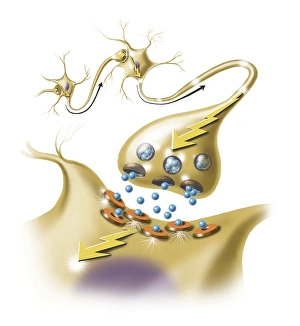
Background imageNeurology Collection: A nerve synapse showing the release of neurotransmitters

A nerve synapse showing the release of neurotransmitters
Detail of a nerve synapse showing the release of neurotransmitters

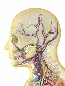
Background imageNeurology Collection: Human face and neck area with nervous system, lymphatic system and circulatory system

Human face and neck area with nervous system, lymphatic system and circulatory system
Human face and neck area with internal throat parts, nervous system, lymphatic system and circulatory system

Background imageNeurology Collection: Microscopic view of nerve fibers

Microscopic view of nerve fibers. A nerve fiber is a threadlike extension of a nerve cell in the nervous system

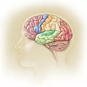
Background imageNeurology Collection: Side view of the human head showing the functional lobes of the brain

Side view of the human head showing the functional lobes of the brain
Side view of the human head showing the functional lobes of the human brain

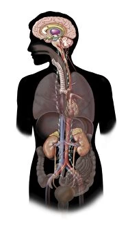
Background imageNeurology Collection: The sympathetic nervous system and the organs of fight-or-flight response

The sympathetic nervous system and the organs of fight-or-flight response

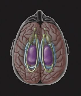
Background imageNeurology Collection: View of limbic system as seen from directly above the head

View of limbic system as seen from directly above the head

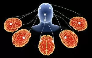
Background imageNeurology Collection: Conceptual image of multi-brain processing

Conceptual image of multi-brain processing

Background imageNeurology Collection: Cross section of brain with arteries

Cross section of brain with arteries

Background imageNeurology Collection: Conceptual image of synapse receptors

Conceptual image of synapse receptors

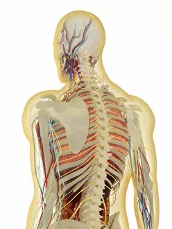
Background imageNeurology Collection: Human body with nervous system, lymphatic system and circulatory system

Human body with nervous system, lymphatic system and circulatory system
Transparent human body with internal organs, nervous system, lymphatic system and circulatory system

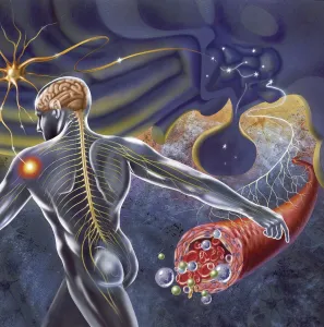
Background imageNeurology Collection: Conceptual image of human nervous system

Conceptual image of human nervous system

Background imageNeurology Collection: Medical illustration showing thyroid cartilage and nerves around neck and head area

Medical illustration showing thyroid cartilage and nerves around neck and head area
Medical illustration showing thyroid cartilage and nerves around human neck and head area

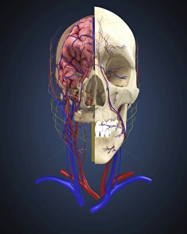
Background imageNeurology Collection: Human skull showing brain and circulatory system

Human skull showing brain and circulatory system

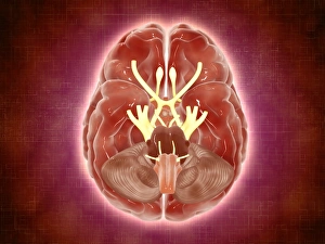
Background imageNeurology Collection: Conceptual image of cranial nerves in human brain

Conceptual image of cranial nerves in human brain

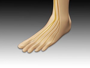
Background imageNeurology Collection: Conceptual image of human foot with nervous system

Conceptual image of human foot with nervous system

Background imageNeurology Collection: Schematic of the hypothalamus receiving nerve impulses from the body

Schematic of the hypothalamus receiving nerve impulses from the body and sending messages to the circulatory and nervous system

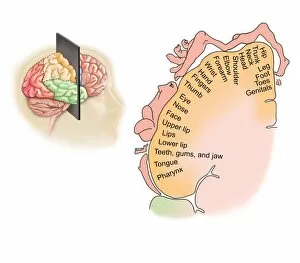
Background imageNeurology Collection: Human brain showing the layout of the sensory cortex

Human brain showing the layout of the sensory cortex
Coronal section through human brain showing the layout of the sensory cortex


All Professionally Made to Order for Quick Shipping

Neurology, the fascinating study of the nervous system, unravels the intricate workings of our brain and its various components. From the Motor homunculus model that depicts how different body parts are represented in our motor cortex to the Histological Diagram of a Mammalian Retina revealing the complexity of our visual processing, every aspect is awe-inspiring. The Cerebellum tissue, as seen through a light micrograph, showcases its unique structure responsible for coordinating movement and balance. Meanwhile, an inferior view of the Anatomy of human brain highlights its remarkable organization and interconnectedness. Through Brain fibres captured by DTI MRI scans like C017 / 7099 or C017 / 7035, we gain insights into neural pathways that facilitate communication between different regions. These pathways form a complex network crucial for transmitting information throughout our brain. Exploring Brain blood vessels using a 3D angiogram (C007 / 1981) reveals their vital role in supplying oxygen and nutrients to sustain neuronal function. Additionally, understanding Motor and sensory homunculi helps us comprehend how specific areas within our brains control different bodily functions. Delving deeper into neurology brings us face-to-face with Medulla oblongata artwork depicting this critical region involved in regulating essential autonomic functions such as breathing and heart rate. Furthermore, observing Nerve and glial cells under a light microscope illustrates their diverse roles in supporting neurons' health and functionality. Finally, examining Synapse nerve junctions through TEM unveils these microscopic structures where electrical signals are transmitted from one neuron to another—a fundamental process underlying all brain activity. In essence, neurology takes us on an incredible journey deep within ourselves—unveiling mysteries at both macroscopic levels like anatomical structures or microscopically exploring cellular intricacies, and is through this exploration that we gain profound knowledge about how our brains work—the very essence of who we are as individuals.