
Lm Collection
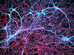
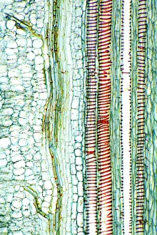
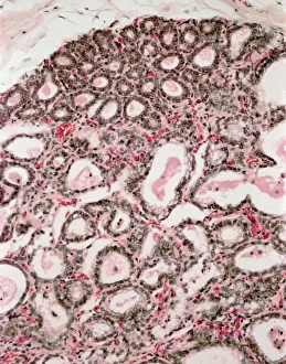
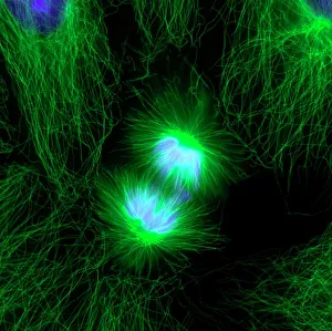
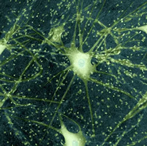
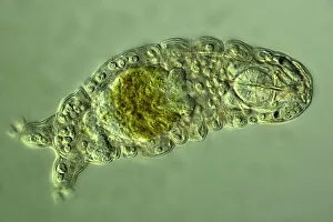
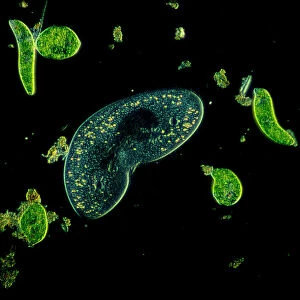
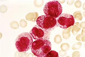
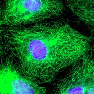
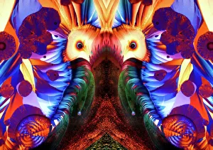
Exploring the intricate network - nerve and glial cells under a light micrograph, revealing the hidden complexities of our brain's functioning


All Professionally Made to Order for Quick Shipping
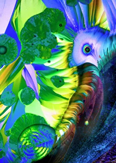
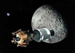
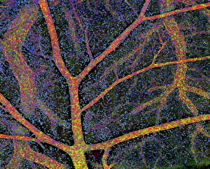
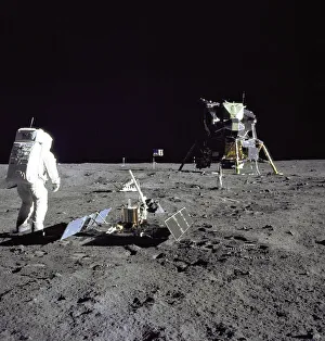
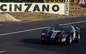
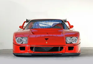
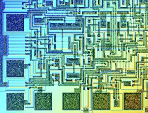
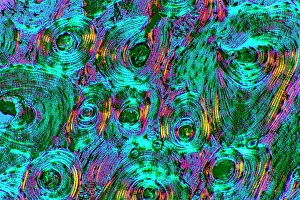
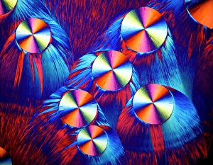
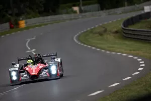
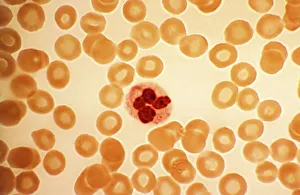
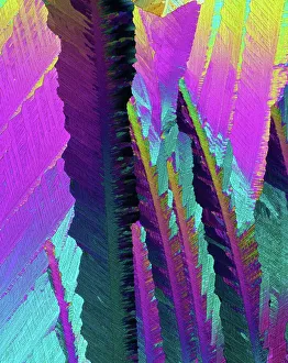
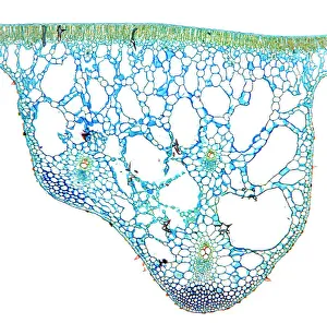
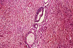
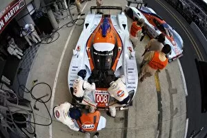
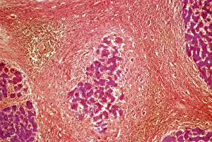
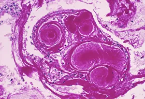
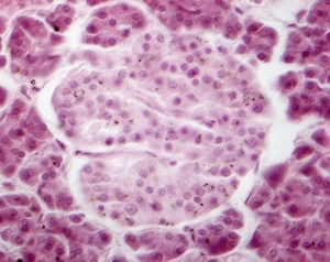
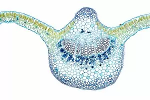
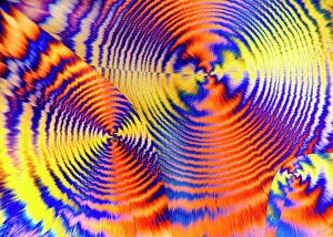
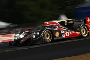
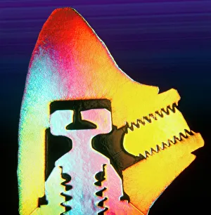
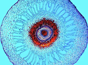
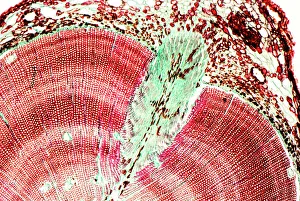
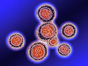
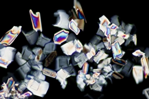
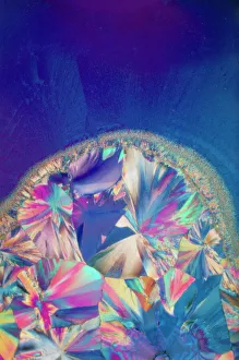
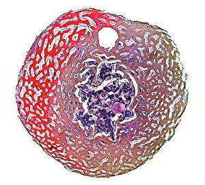
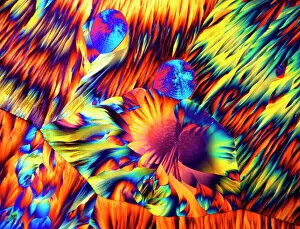
Exploring the intricate network - nerve and glial cells under a light micrograph, revealing the hidden complexities of our brain's functioning. The stunning combination of copper and magnesium sulphate in LM creates a mesmerizing visual display reminiscent of an Apollo spacecraft at the Moon, captured through artistic interpretation. Revving up with power and speed, the Ferrari F40 LM takes center stage, showcasing its dominance on the racetrack. Delving deeper into nature's wonders, we uncover the delicate structure of a dicotyledon plant stem under a light micrograph, highlighting its resilience and growth potential. Buzz Aldrin's nostalgic gaze back at Tranquility Base reminds us of humanity's remarkable achievements in space exploration. Reliving history, we transport ourselves to Le Mans 24 Hours in France on June 19th, 1966 - an iconic race that forever etched its mark on motorsport history. Fast forward to present day excitement as we witness the thrilling action unfold at the 2018 24 Hours of Le Mans. Peering into our body's vital systems, we discover how blood supply nourishes our brain tissue for optimal function and health. Embracing Italian excellence once again with Ferrari F40 LM Italy edition - where style meets performance in automotive perfection. Shedding light on motherhood's miracles, lactating breast tissue reveals its unique beauty under a light micrograph - symbolizing life-giving sustenance for newborns. Unveiling technology's marvels from within, a microscopic view captures intricate details of a microchip - showcasing innovation that powers our modern world.