Fraud Blocker Skip to main content

Immunology Collection (page 5)

"Unleashing the Power of Immunology: Exploring the Intricate World of Immune Responses" Immunology

Background imageImmunology Collection: Human antitumour antibody molecule

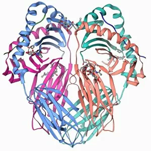
Human antitumour antibody molecule
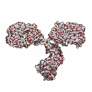
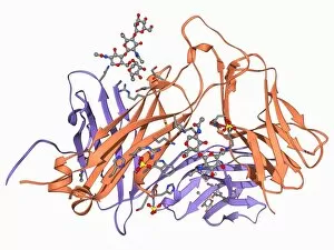
Human antitumour antibody. Molecular model showing the antitumour antibody BR96 complexed with part of the Lewis antigen. The Lewis antigen is expressed on the surface of human carcinoma cells

Background imageImmunology Collection: Edward Jenner, British physician C017 / 7108

Edward Jenner, British physician C017 / 7108
Edward Jenner (1749-1823), British physician. Jenner, who also did work as a naturalist, is famed for developing a vaccine for the often fatal viral infection smallpox (syringe in hand)

Background imageImmunology Collection: Antibody molecule F007 / 0109

Antibody molecule F007 / 0109
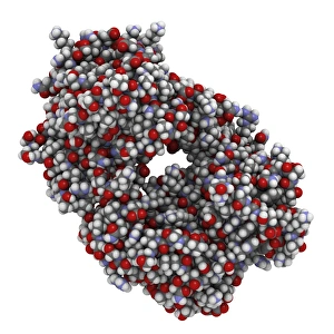
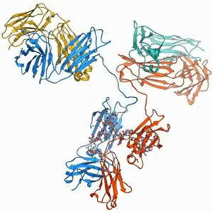
Antibody molecule. Crystal structure of a monoclonal immunoglobulin (IgG2a). IgG antibodies are composed of 2 long heavy chains and 2 shorter light chains

Background imageImmunology Collection: Antibody molecule F007 / 0107

Antibody molecule F007 / 0107
Antibody molecule. Crystal structure of a monoclonal immunoglobulin (IgG2a). IgG antibodies are composed of 2 long heavy chains and 2 shorter light chains

Background imageImmunology Collection: Antibody molecule F007 / 0104

Antibody molecule F007 / 0104
Antibody molecule. Crystal structure of a monoclonal immunoglobulin (IgG2a). IgG antibodies are composed of 2 long heavy chains and 2 shorter light chains

Background imageImmunology Collection: Antibody molecule F007 / 0105

Antibody molecule F007 / 0105
Antibody molecule. Crystal structure of a monoclonal immunoglobulin (IgG2a). IgG antibodies are composed of 2 long heavy chains and 2 shorter light chains

Background imageImmunology Collection: Antibody molecule F007 / 0103

Antibody molecule F007 / 0103
Antibody molecule. Crystal structure of a monoclonal immunoglobulin (IgG2a). IgG antibodies are composed of 2 long heavy chains and 2 shorter light chains

Background imageImmunology Collection: Alemtuzumab Fab fragment molecule F007 / 0100

Alemtuzumab Fab fragment molecule F007 / 0100
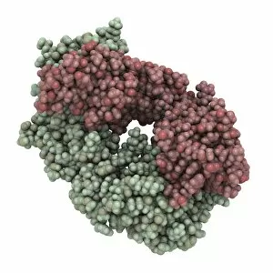
Alemtuzumab Fab fragment, crystal structure. Alemtuzumab is a humanized monoclonal antibody that binds the CD52 protein and is used in the treatment of cancer and auto-immune disease

Background imageImmunology Collection: Antibody molecule F007 / 0102

Antibody molecule F007 / 0102
Antibody molecule. Crystal structure of a monoclonal immunoglobulin (IgG2a). IgG antibodies are composed of 2 long heavy chains and 2 shorter light chains

Background imageImmunology Collection: Alemtuzumab Fab fragment molecule F007 / 0099

Alemtuzumab Fab fragment molecule F007 / 0099
Alemtuzumab Fab fragment, crystal structure. Alemtuzumab is a humanized monoclonal antibody that binds the CD52 protein and is used in the treatment of cancer and auto-immune disease

Background imageImmunology Collection: Antibody molecule F007 / 0101

Antibody molecule F007 / 0101
Antibody molecule. Crystal structure of a monoclonal immunoglobulin (IgG2a). IgG antibodies are composed of 2 long heavy chains and 2 shorter light chains

Background imageImmunology Collection: Alemtuzumab Fab fragment molecule F007 / 0097

Alemtuzumab Fab fragment molecule F007 / 0097
Alemtuzumab Fab fragment, crystal structure. Alemtuzumab is a humanized monoclonal antibody that binds the CD52 protein and is used in the treatment of cancer and auto-immune disease

Background imageImmunology Collection: Alemtuzumab Fab fragment molecule F007 / 0098

Alemtuzumab Fab fragment molecule F007 / 0098
Alemtuzumab Fab fragment, crystal structure. Alemtuzumab is a humanized monoclonal antibody that binds the CD52 protein and is used in the treatment of cancer and auto-immune disease

Background imageImmunology Collection: White blood cell and platelet, SEM F006 / 9929

White blood cell and platelet, SEM F006 / 9929
White blood cells. Coloured scanning electron micrograph (SEM) of white blood cells (leucocytes) and a platelet (thrombocyte, red). Magnification: x6, 600 when printed at 10 centimetres wide
Background imageImmunology Collection: White blood cell and platelet, SEM F006 / 9928

White blood cell and platelet, SEM F006 / 9928
White blood cells. Coloured scanning electron micrograph (SEM) of white blood cells (leucocytes) and a platelet (thrombocyte, orange). Magnification: x6, 600 when printed at 10 centimetres wide

Background imageImmunology Collection: White blood cell and platelet, SEM F006 / 9927

White blood cell and platelet, SEM F006 / 9927
White blood cells. Coloured scanning electron micrograph (SEM) of white blood cells (leucocytes) and a platelet (thrombocyte, green). Magnification: x6, 600 when printed at 10 centimetres wide

Background imageImmunology Collection: White blood cells, SEM F006 / 9924

White blood cells, SEM F006 / 9924
White blood cells. Coloured scanning electron micrograph (SEM) of white blood cells (leucocytes). Magnification: x2, 400 when printed at 10 centimetres wide

Background imageImmunology Collection: White blood cells, SEM F006 / 9923

White blood cells, SEM F006 / 9923
White blood cells. Coloured scanning electron micrograph (SEM) of white blood cells (leucocytes). Magnification: x2, 400 when printed at 10 centimetres wide

Background imageImmunology Collection: White blood cells, SEM F006 / 9926

White blood cells, SEM F006 / 9926
White blood cells. Coloured scanning electron micrograph (SEM) of white blood cells (leucocytes). Magnification: x2, 400 when printed at 10 centimetres wide

Background imageImmunology Collection: White blood cells, SEM F006 / 9925

White blood cells, SEM F006 / 9925
White blood cells. Coloured scanning electron micrograph (SEM) of white blood cells (leucocytes). Magnification: x2, 400 when printed at 10 centimetres wide

Background imageImmunology Collection: White blood cells, SEM F006 / 9922

White blood cells, SEM F006 / 9922
White blood cells. Coloured scanning electron micrograph (SEM) of white blood cells (leucocytes). Magnification: x2, 400 when printed at 10 centimetres wide

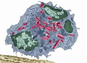
Background imageImmunology Collection: Eosinophil white blood cell, TEM C014 / 1437

Eosinophil white blood cell, TEM C014 / 1437
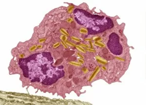
Eosinophil white blood cell. Transmission electron micrograph (TEM) of a section through an eosinophil. Eosinophils, like all white blood cells, are part of the bodys immune system

Background imageImmunology Collection: Activated macrophage, SEM F006 / 8635

Activated macrophage, SEM F006 / 8635
Activated human macrophage, coloured scanning electron micrograph (SEM). Magnification: x2, 700 when printed at 10 centimetres wide

Background imageImmunology Collection: Activated macrophage, SEM F006 / 8634

Activated macrophage, SEM F006 / 8634
Activated human macrophage, coloured scanning electron micrograph (SEM). Magnification: x2, 700 when printed at 10 centimetres wide

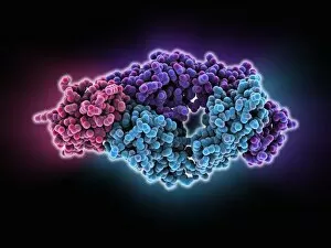
Background imageImmunology Collection: Antibodies and their antigen F006 / 9678

Antibodies and their antigen F006 / 9678
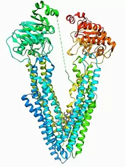
Antibodies and their antigen. Molecular model showing the molecular structure of two anti-P-glycoprotein antibodies (upper left and right) and the P-glycoprotein (v-shaped) to which they respond

Background imageImmunology Collection: Toll-like receptor 3 and RNA F006 / 9666

Toll-like receptor 3 and RNA F006 / 9666
Toll-like receptor 3 and RNA. Molecular model of the toll-like receptor 3 (TLR3) protein (pink and blue) bound to a strand of RNA (ribonucleic acid, green and yellow)

Background imageImmunology Collection: Interferon antagonism by viral protein F006 / 9662

Interferon antagonism by viral protein F006 / 9662
Interferon (IFN) antagonism by viral protein. Molecular model of an orthopoxvirus IFN-gamma-binding protein bound to an IFN-gamma molecule

Background imageImmunology Collection: HIV antibody therapy, molecular model F006 / 9622

HIV antibody therapy, molecular model F006 / 9622
HIV antibody therapy. Molecular model of the interaction of the HIV surface protein gp120 (green) as it interacts with a human white blood cell surface protein (CD4)

Background imageImmunology Collection: T cell receptor, molecular model F006 / 9515

T cell receptor, molecular model F006 / 9515
T cell receptor. Molecular model of an alpha T cell receptor. T cell receptors are protein complexes found on the surface of a type of white blood cell called T lymphocytes (or T cells)

Background imageImmunology Collection: Flu virus surface protein and antibody F006 / 9428

Flu virus surface protein and antibody F006 / 9428
Flu virus surface protein and antibody. Molecular model of the neuraminidase glycoprotein enzyme found on the surface of the influenza (flu) virus

Background imageImmunology Collection: Immunoglobulin G antibody molecule F006 / 9371

Immunoglobulin G antibody molecule F006 / 9371
Immunoglobulin G (IgG) antibody, molecular model. This is the most abundant immunoglobulin and is found in all body fluids

Background imageImmunology Collection: Human interferon alpha molecule F006 / 9373

Human interferon alpha molecule F006 / 9373
Human interferon alpha, molecular model. Interferons are proteins produced by white blood cells as part of the immune response to invading pathogens, especially viruses

Background imageImmunology Collection: MHC protein-antigen complex F006 / 9363

MHC protein-antigen complex F006 / 9363
MHC protein-antigen complex. Molecular model of the human class I MHC (major histocompatibility complex) protein HLA-B27 complexed with beta-2 microglobulin

Background imageImmunology Collection: Eosinophil white blood cell, TEM C014 / 1439

Eosinophil white blood cell, TEM C014 / 1439
Eosinophil white blood cell. Transmission electron micrograph (TEM) of a section through an eosinophil. Eosinophils, like all white blood cells, are part of the bodys immune system

Background imageImmunology Collection: T cell receptor antigen complex F006 / 9339

T cell receptor antigen complex F006 / 9339
T cell receptor antigen complex. Molecular model of the alphabeta T cell receptor bound to the influenza haemagglutinin antigen and MHC class II molecule HLA-DR1

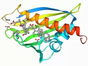
Background imageImmunology Collection: Birch pollen allergen F006 / 9335

Birch pollen allergen F006 / 9335
Birch pollen allergen. Molecular model of Bet v 1l, the molecule responsible for allergic reactions to birch pollen. Here

Background imageImmunology Collection: MHC protein complexed with flu virus F006 / 9294

MHC protein complexed with flu virus F006 / 9294
MHC protein complexed with flu virus. Molecular model showing human class II MHC (major histocompatibility complex) protein HLA-DR1 complexed with an influenza (flu) virus peptide

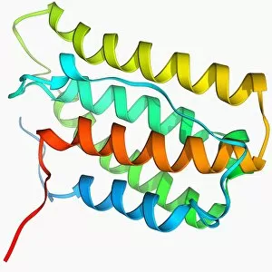
Background imageImmunology Collection: CD4 protein molecule F006 / 9266

CD4 protein molecule F006 / 9266
CD4 protein. Molecular model of the CD4 T cell (T lymphocyte) receptor protein. CD4 binds to the surface proteins on other cells and pathogenic micro-organisms

Background imageImmunology Collection: T cell receptor B7 molecule F006 / 9247

T cell receptor B7 molecule F006 / 9247
T cell receptor B7. Molecular model of the T cell receptor B7 bound to the viral Tax peptide and MHC class I molecule HLA-A2

Background imageImmunology Collection: Human interferon beta molecule F006 / 9241

Human interferon beta molecule F006 / 9241
Human interferon beta, molecular model. Interferons are proteins produced by white blood cells as part of the immune response to invading pathogens, especially viruses

Background imageImmunology Collection: Interleukin-6, molecular model F006 / 9232

Interleukin-6, molecular model F006 / 9232
Interleukin-6. Molecular model of the cytokine protein human interleukin-6. This protein is produced in the body and has a wide variety of functions in the immune system

Background imageImmunology Collection: HIV antibody therapy, molecular model C018 / 9193

HIV antibody therapy, molecular model C018 / 9193
HIV antibody therapy. 3D model of the interaction of the HIV surface protein gp120 as it interacts with a human white blood cell surface protein (CD4) and the anti-HIV antibody (17b)

Background imageImmunology Collection: Antibodies and viruses, artwork

Antibodies and viruses, artwork
Antibodies and viruses. Computer artwork showing antibody, or immunoglobulin, molecules (blue) surrounding virus particles (virions, yellow)

Background imageImmunology Collection: T-cell receptor bound to enterotoxin

T-cell receptor bound to enterotoxin, molecular model. The T cell receptor (TCR) is a protein complex found on the surface of a type of white blood cell called T lymphocytes (or T cells)

Background imageImmunology Collection: NK cells attacking cancer cells, artwork

NK cells attacking cancer cells, artwork
NK cells attacking cancer cells. Computer artwork of human Natural Killer (NK) cells (small, round) attacking cancer cells (large, round)

Background imageImmunology Collection: Immunotherapy, artwork

Immunotherapy, artwork
Immunotherapy. Computer artwork of immunotherapy being used to destroy a tumour (right). Immunotherapy uses the bodys immune system to fight a disease

Background imageImmunology Collection: Grass pollen allergen molecule

Grass pollen allergen molecule
Grass pollen allergen. Molecular model of the major grass pollen allergen Phl p2 complexed with the antigen-binding fragment (fab) of its human immunoglobulin E antibody

Background imageImmunology Collection: Antibodies and bacteria, artwork

Antibodies and bacteria, artwork
Antibodies and bacteria. Computer artwork showing white blood cells (large, round) and antibody, or immunoglobulin, molecules (red) surrounding bacteria (green)


All Professionally Made to Order for Quick Shipping

"Unleashing the Power of Immunology: Exploring the Intricate World of Immune Responses" Immunology, a fascinating field that delves into the complex mechanisms of our immune system, holds immense potential in combating diseases. T lymphocytes and cancer cells engage in a constant battle for supremacy, as depicted by SEM C001 / 1679. These tiny warriors play a crucial role in identifying and eliminating abnormal cells. The Immunoglobulin G antibody molecule (F007 / 9894) stands tall as one of our body's most powerful defenders against pathogens. Its remarkable structure enables it to neutralize harmful invaders with precision and efficiency. Neutrophils, exemplified by SEM C018 / 8596, showcase their extraordinary ability to engulf MRSA bacteria—an awe-inspiring sight indeed. Meanwhile, dendritic cells (artwork) act as vigilant sentinels, capturing antigens and presenting them to other immune cells for recognition. TEM reveals an up-close view of human white blood cells bearing HLA antigens—a key component in distinguishing self from non-self. Antibodies (artwork), resembling elegant warriors on a mission, bind specifically to foreign substances to mark them for destruction. HIV reverse transcription enzyme serves as a reminder of the challenges faced by immunologists worldwide. This relentless virus exploits our own cellular machinery but continues to be targeted through innovative research efforts. Human macrophages (TEM) demonstrate their exceptional phagocytic abilities while basophil white blood cells stand ready at the frontlines—both integral players in mounting effective immune responses against invading pathogens. Intriguingly captured by SEM imagery is bacteria infecting a macrophage—a visual representation highlighting how these microscopic organisms can exploit host defenses while also serving as valuable tools for studying infection dynamics. Lastly, Dohle bodies within blood cells offer insights into various pathological conditions affecting neutrophils—an essential clue guiding immunologists towards understanding and treating immune disorders.