Fraud Blocker Skip to main content

Immunology Collection (page 2)

"Unleashing the Power of Immunology: Exploring the Intricate World of Immune Responses" Immunology

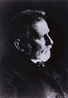
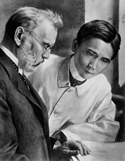
Background imageImmunology Collection: Paul Ehrlich, German doctor and scientist (b / w photo)

Paul Ehrlich, German doctor and scientist (b / w photo)
6014954 Paul Ehrlich, German doctor and scientist (b/w photo) by German Photographer (20th Century); Private Collection; (add.info.: Paul Ehrlich (1854-1915)

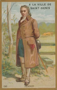
Background imageImmunology Collection: Edward Jenner, English doctor and discoverer of a vaccine for smallpox. (chromolitho)

Edward Jenner, English doctor and discoverer of a vaccine for smallpox. (chromolitho)
668844 Edward Jenner, English doctor and discoverer of a vaccine for smallpox. (chromolitho) by French School, (19th century); Private Collection; (add.info.: Edward Jenner)

Background imageImmunology Collection: Louis Pasteur and his collaborators in working on a vaccine for croup at the Institut Pasteur in

Louis Pasteur and his collaborators in working on a vaccine for croup at the Institut Pasteur in Paris (b / w photo)
3098643 Louis Pasteur and his collaborators in working on a vaccine for croup at the Institut Pasteur in Paris (b/w photo) by French Photographer

Background imageImmunology Collection: Peaceful glories of France (colour litho)

Peaceful glories of France (colour litho)
3099324 Peaceful glories of France (colour litho) by French School, (19th century); Private Collection; (add.info.: Peaceful glories of France)

Background imageImmunology Collection: Jean-Baptiste Jupille, French shepherd boy who fought off a rabid dog to prevent it attacking

Jean-Baptiste Jupille, French shepherd boy who fought off a rabid dog to prevent it attacking other children
974398 Jean-Baptiste Jupille, French shepherd boy who fought off a rabid dog to prevent it attacking other children (chromolitho) by European School

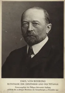
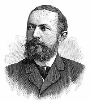
Background imageImmunology Collection: Emil von Behring, German doctor and physiologist, c1890 (b / w photo)

Emil von Behring, German doctor and physiologist, c1890 (b / w photo)
5232155 Emil von Behring, German doctor and physiologist, c1890 (b/w photo) by German Photographer, (19th century); Private Collection; (add.info.: Emil von Behring (1854-1917)

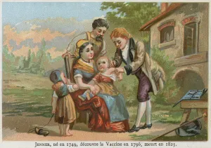
Background imageImmunology Collection: Edward Jenner (chromolitho)

Edward Jenner (chromolitho)
961327 Edward Jenner (chromolitho) by English School, (19th century); Private Collection; (add.info.: Edward Jenner, born in 1749, discovered the vaccine in 1796, died in 1823)

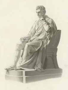
Background imageImmunology Collection: Edward Jenner, English physician and scientist (engraving)

Edward Jenner, English physician and scientist (engraving)
979428 Edward Jenner, English physician and scientist (engraving) by Marshall, William Calder (1813-94); Private Collection; (add.info.: Edward Jenner (1749-1823), English physician and scientist

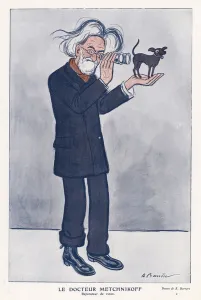
Background imageImmunology Collection: Elie Metchnikoff, Russian zoologist (colour litho)

Elie Metchnikoff, Russian zoologist (colour litho)
3620555 Elie Metchnikoff, Russian zoologist (colour litho) by Barrere, Adrien (1877-1931); Private Collection; (add.info.: Elie Metchnikoff (1845-1916), Russian zoologist.); © Look and Learn

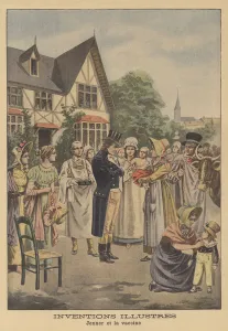
Background imageImmunology Collection: Edward Jenners smallpox vaccine (colour litho)

Edward Jenners smallpox vaccine (colour litho)
2819130 Edward Jenners smallpox vaccine (colour litho) by French School, (20th century); Private Collection; (add.info.: Edward Jenners smallpox vaccine. Inventions illustres. Jenner et la vaccine)
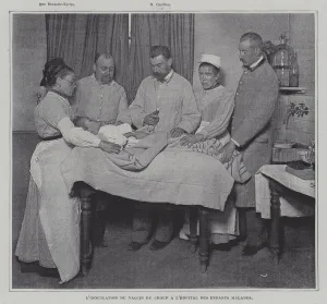
Background imageImmunology Collection: Inoculation of a patient with the vaccine against croup at a hospital for sick children in Paris

Inoculation of a patient with the vaccine against croup at a hospital for sick children in Paris (b / w photo)
3098642 Inoculation of a patient with the vaccine against croup at a hospital for sick children in Paris (b/w photo) by French Photographer

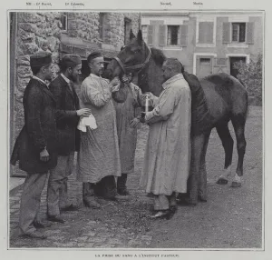
Background imageImmunology Collection: Taking a sample of blood from a horse at the Institut Pasteur, Paris (b / w photo)

Taking a sample of blood from a horse at the Institut Pasteur, Paris (b / w photo)
3098645 Taking a sample of blood from a horse at the Institut Pasteur, Paris (b/w photo) by French Photographer, (19th century); Private Collection; (add.info)

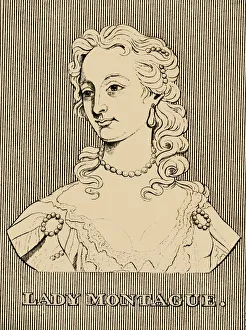
Background imageImmunology Collection: Lady Montague, (1689- 1762), 1830. Creator: Unknown
Lady Montague, (1689- 1762), 1830. Creator: Unknown
Lady Montague, (1689- 1762), 1830. Lady Mary Wortley Montague (1689- 1762) English aristocrat, letter writer, and poet, who wrote during travels to the Ottoman Empire

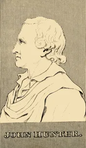
Background imageImmunology Collection: John Hunter, (1728-1793), 1830. Creator: Unknown

John Hunter, (1728-1793), 1830. Creator: Unknown
John Hunter, (1728-1793), 1830. John Hunter (1728-1793) Scottish surgeon and scientist, pioneer of the smallpox vaccine and expert in anatomy who became a Fellow of the Royal Society in 1767

Background imageImmunology Collection: Edward Jenner M. D. (1749-1823), 1830. Creator: Unknown

Edward Jenner M. D. (1749-1823), 1830. Creator: Unknown
Edward Jenner M.D. (1749-1823), 1830. Edward Jenner (1749-1823) English physician and scientist, pioneer of smallpox vaccine, known as the father of immunology

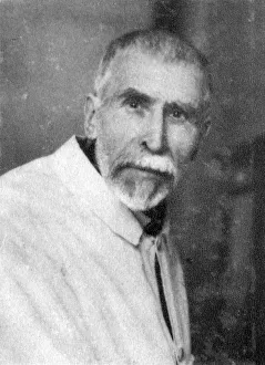
Background imageImmunology Collection: Pierre Paul Emile Roux, French physician, bacteriologist and immunologist, 1928

Pierre Paul Emile Roux, French physician, bacteriologist and immunologist, 1928. Roux (1853-1933) was one of the closest collaborators of Louis Pasteur

Background imageImmunology Collection: Ilya Ilyich Mechnikov, Russian microbiologist, c1900s(?). Artist: Nadar

Ilya Ilyich Mechnikov, Russian microbiologist, c1900s(?). Artist: Nadar
Ilya Ilyich Mechnikov, Russian microbiologist, c1900s(?). Mechnikov (1845-1916) continued the work of Louis Pasteur, specialising in the study of the immune system

Background imageImmunology Collection: Emil von Behring, German immunologist and bacteriologist, 1902

Emil von Behring, German immunologist and bacteriologist, 1902. In 1901 von Behring (1854-1917) was awarded the first Nobel Prize for Physiology or Medicine for his development of a serum for

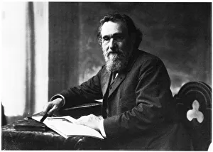
Background imageImmunology Collection: Ilya Ilich Mechnikov, Russian microbiologist

Ilya Ilich Mechnikov, Russian microbiologist. Mechnikov (1845-1916) continued the work of Louis Pasteur, specialising in the study of the immune system

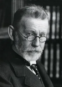
Background imageImmunology Collection: Paul Ehrlich (1854-1915), German bacteriologist

Paul Ehrlich (1854-1915), German bacteriologist
Paul Ehrlich (1854-1915) German bacteriologist. Specialist in the fields of Haematology, Chemotherapy and Immunology. Shared Nobel prize for medicine or physiology with Mechnikov in 1908

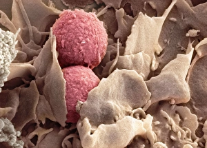
Background imageImmunology Collection: Phagocytosis of fungus spores, SEM

Phagocytosis of fungus spores, SEM
Phagocytosis of fungus spores. Coloured scanning electron micrograph (SEM) of spores from the fungus Aspergillus fumigatus (red) being phagocytosed (engulfed and destroyed) by a dendritic cell (brown)

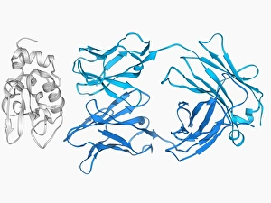
Background imageImmunology Collection: Immunoglobulin G antibody and egg white F006 / 9682

Immunoglobulin G antibody and egg white F006 / 9682
Immunoglobulin G and egg white. Molecular model of an immunoglobulin G (IgG) antibody bound to a molecule of egg white. This is the most abundant immunoglobulin and is found in all body fluids

Background imageImmunology Collection: Thymus gland, light micrograph C015 / 4970

Thymus gland, light micrograph C015 / 4970
Thymus gland. Light micrograph of a section through tissue from the thymus gland, part of the lymphoid system. The dense areas are the thymic cortex, which produces lots of lymphocytes

Background imageImmunology Collection: Myeloblast blood cell, light micrograph

Myeloblast blood cell, light micrograph
Myeloblast blood cell. Light micrograph of blood cells, including a myeloblast, a precursor for a type of white blood cell (leucocyte). Myeloblasts differentiate into granulocytes

Background imageImmunology Collection: Eosinophil white blood cell, TEM

Eosinophil white blood cell, TEM
Eosinophil white blood cell. Transmission electron micrograph (TEM) of a section through an eosinophil white blood cell (leukocyte)

Background imageImmunology Collection: Promyelocyte blood cell, light micrograph

Promyelocyte blood cell, light micrograph. This blood cell (centre) is a precursor for a type of white blood cell called a granulocyte, formed by granulopoiesis in the bone marrow

Background imageImmunology Collection: Chronic lymphocytic leukaemia, micrograph

Chronic lymphocytic leukaemia, micrograph
Chronic lymphocytic leukaemia. Light micrograph of blood cells sampled from a lymphatic ganglion in a case of chronic lymphocytic leukaemia, also called chronic lymphoid leukaemia (CLL)

Background imageImmunology Collection: Macrophage and lymphocytes, TEM

Macrophage and lymphocytes, TEM
Macrophage and lymphocytes, transmission electron micrograph (TEM). Macrophage surrounded by lymphocytes in a lymph node. This view includes many intercellular contacts

Background imageImmunology Collection: Immunoglobulin A, molecular model

Immunoglobulin A, molecular model
Immunoglobulin A. Molecular model of the structure of the antibody immunoglobulin A (IgA). This is the secretory dimeric form (sIgA), the main immunoglobulin found in secretions such as saliva

Background imageImmunology Collection: Immunoglobulin G antibody molecule C016 / 4462

Immunoglobulin G antibody molecule C016 / 4462
Immunoglobulin G antibody molecule. Computer artwork of a model of the secondary structure of immunoglobulin G (IgG). This is the most abundant immunoglobulin and is found in all body fluids

Background imageImmunology Collection: White blood cells and platelets, SEM C016 / 3099

White blood cells and platelets, SEM C016 / 3099
White blood cells and platelets. Coloured scanning electron micrograph (SEM) of platelets (green) with a variety of different white blood cells (leucocytes, yellow)

Background imageImmunology Collection: White blood cells and platelets, SEM C016 / 3098

White blood cells and platelets, SEM C016 / 3098
White blood cells and platelets. Coloured scanning electron micrograph (SEM) of platelets (purple) with a variety of different white blood cells (leucocytes, blue)

Background imageImmunology Collection: Monocyte white blood cell, SEM C016 / 3089

Monocyte white blood cell, SEM C016 / 3089
Monocyte white blood cell. Scanning electron micrograph (SEM) of a monocyte white blood cell. Monocytes, like all white blood cells, are part of the human bodys immune system

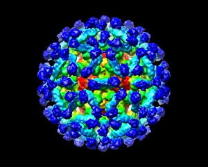
Background imageImmunology Collection: Murine norovirus with antibody fragments

Murine norovirus with antibody fragments
Murine norovirus (MNV) with antibody fragments, computer model. This image was created using molecular modelling software and data from cryo- electron microscopy

Background imageImmunology Collection: EHRLICH, Paul (1854-1915). German physician

EHRLICH, Paul (1854-1915). German physician and scientist who worked in the fields of hematology, immunology, and chemotherapy. Nobel Prize in 1908. Engraving

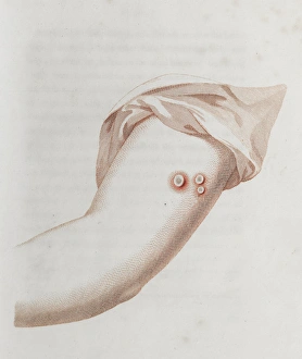
Background imageImmunology Collection: Coloured depiction of lesion on arm

Coloured depiction of lesion on arm Source: An inquiry into the causes and effects of the variol栶 accinae, a disease discovered in some of the western counties of England

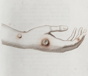
Background imageImmunology Collection: Coloured depiction of lesion on a hand

Coloured depiction of lesion on a hand Source: An inquiry into the causes and effects of the variol栶 accinae, a disease discovered in some of the western counties of England

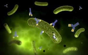
Background imageImmunology Collection: Conceptual image of antibody attaching and killing bacteria

Conceptual image of antibody attaching and killing bacteria

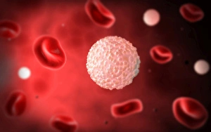
Background imageImmunology Collection: Microscopic view of white blood cells inside blood vessel

Microscopic view of white blood cells inside blood vessel

Background imageImmunology Collection: 3D rendering of human circulatory system

3D rendering of human circulatory system

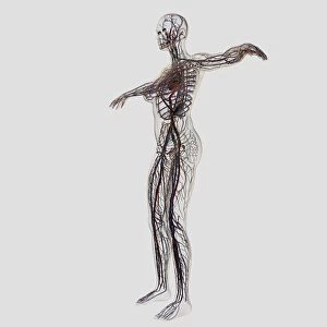
Background imageImmunology Collection: Medical illustration of female arteries, veins and lymphatic system

Medical illustration of female arteries, veins and lymphatic system, full body view

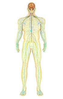
Background imageImmunology Collection: Anatomy of human nervous system and lymphatic system

Anatomy of human nervous system and lymphatic system, front view

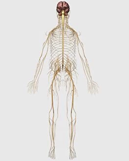
Background imageImmunology Collection: Medical illustration of peripheral nervous system with brain

Medical illustration of peripheral nervous system with brain

Background imageImmunology Collection: 3D rendering of human skull with lymphatic system

3D rendering of human skull with lymphatic system, side view

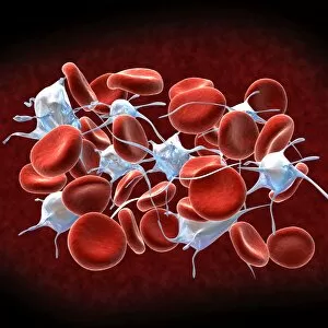
Background imageImmunology Collection: Red blood cells with leukocytes

Red blood cells with leukocytes

Background imageImmunology Collection: Conceptual image of an antibody

Conceptual image of an antibody

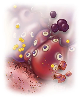
Background imageImmunology Collection: Surrounding tissue becomes inflamed causing several systemic effects

Surrounding tissue becomes inflamed causing several systemic effects, such as vasodilation, mucous secretion, nerve stimulation and smooth muscle contraction

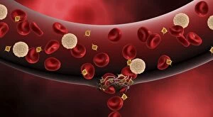
Background imageImmunology Collection: Blood vessel with platelets, white blood cells and red blood cells

Blood vessel with platelets, white blood cells and red blood cells


All Professionally Made to Order for Quick Shipping

"Unleashing the Power of Immunology: Exploring the Intricate World of Immune Responses" Immunology, a fascinating field that delves into the complex mechanisms of our immune system, holds immense potential in combating diseases. T lymphocytes and cancer cells engage in a constant battle for supremacy, as depicted by SEM C001 / 1679. These tiny warriors play a crucial role in identifying and eliminating abnormal cells. The Immunoglobulin G antibody molecule (F007 / 9894) stands tall as one of our body's most powerful defenders against pathogens. Its remarkable structure enables it to neutralize harmful invaders with precision and efficiency. Neutrophils, exemplified by SEM C018 / 8596, showcase their extraordinary ability to engulf MRSA bacteria—an awe-inspiring sight indeed. Meanwhile, dendritic cells (artwork) act as vigilant sentinels, capturing antigens and presenting them to other immune cells for recognition. TEM reveals an up-close view of human white blood cells bearing HLA antigens—a key component in distinguishing self from non-self. Antibodies (artwork), resembling elegant warriors on a mission, bind specifically to foreign substances to mark them for destruction. HIV reverse transcription enzyme serves as a reminder of the challenges faced by immunologists worldwide. This relentless virus exploits our own cellular machinery but continues to be targeted through innovative research efforts. Human macrophages (TEM) demonstrate their exceptional phagocytic abilities while basophil white blood cells stand ready at the frontlines—both integral players in mounting effective immune responses against invading pathogens. Intriguingly captured by SEM imagery is bacteria infecting a macrophage—a visual representation highlighting how these microscopic organisms can exploit host defenses while also serving as valuable tools for studying infection dynamics. Lastly, Dohle bodies within blood cells offer insights into various pathological conditions affecting neutrophils—an essential clue guiding immunologists towards understanding and treating immune disorders.