
Immunofluorescence Collection
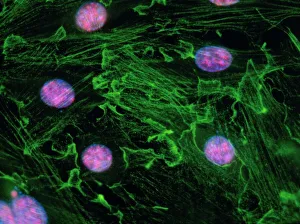
Immunofluorescence is a powerful technique used in the field of biology to visualize and study various cell structures and processes


All Professionally Made to Order for Quick Shipping
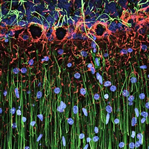
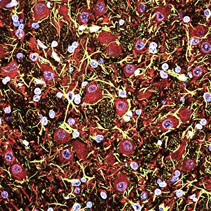
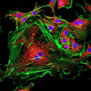
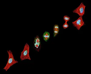
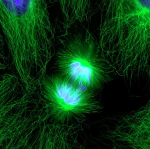
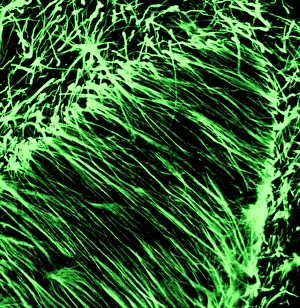
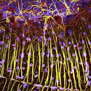
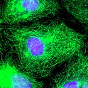
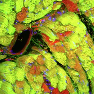
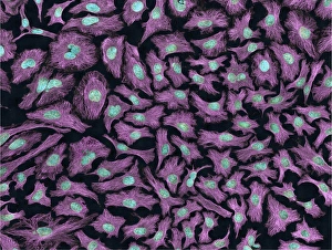
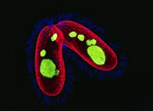
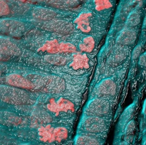
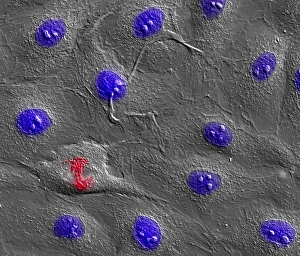
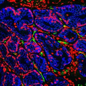
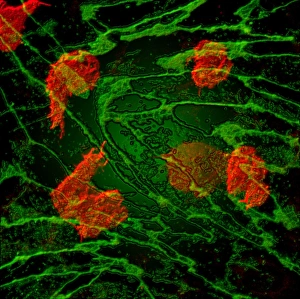
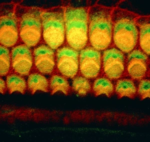
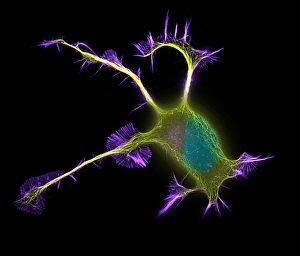
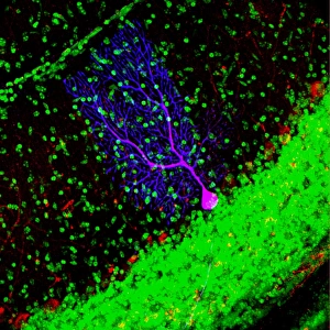
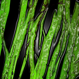
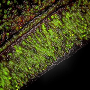
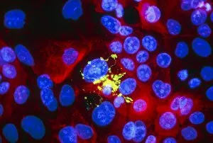
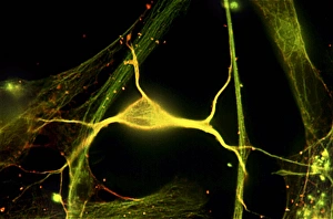
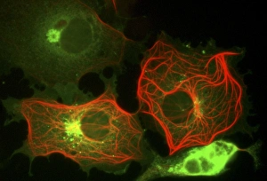
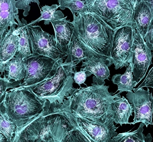
Immunofluorescence is a powerful technique used in the field of biology to visualize and study various cell structures and processes. In this captivating collection of images, we delve into the intricate world of immunofluorescence. Starting with a light micrograph of cerebellum tissue, we are instantly drawn into the mesmerizing beauty of cell structure. The delicate network of neurons and glial cells within the cerebellum comes alive under fluorescent lighting, revealing their vibrant colors and interconnectedness. Moving on to another stunning image, we witness mitosis captured through a light micrograph. This dynamic process showcases the division of cells as they multiply and regenerate. The bright fluorescence highlights each stage with precision, leaving us in awe of nature's remarkable ability to perpetuate life. Shifting our focus to cerebral cortex nerve cells, we explore their intricate architecture through immunofluorescent imaging. These specialized cells play a crucial role in cognition and sensory perception, making them an intriguing subject for scientific investigation. The journey continues with another fluorescent micrograph capturing cell division once again but this time showcasing its ethereal beauty within cerebellum tissue. The glowing hues emphasize the complexity and elegance inherent in cellular replication – an essential process for growth and repair throughout our bodies. Our exploration takes an unexpected turn as we observe uterine cells during childbirth using immunofluorescence microscopy. This extraordinary image captures the intense activity occurring at this pivotal moment when new life enters the world - a testament to both strength and vulnerability simultaneously displayed by these incredible cells. Concluding our visual odyssey is a confocal light micrograph featuring heart muscle tissue. With its rhythmic contractions vital for sustaining life, it is no wonder that studying cardiac cells has always fascinated scientists. Through immunofluorescence techniques, we gain insight into their unique structure while appreciating their intrinsic beauty from every angle. Immunofluorescence allows us to unlock secrets hidden within the microscopic world, revealing the wonders of cell structure and function.