Immunofluorescent LM of fibroblast cell nuclei

![]()
Wall Art and Photo Gifts from Science Photo Library
Immunofluorescent LM of fibroblast cell nuclei
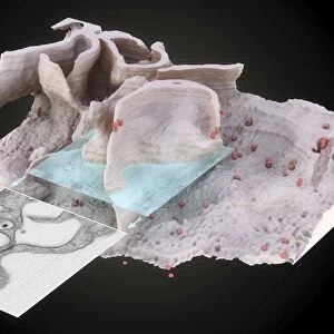
Cell nuclei. Immunofluorescent light micrograph of cultured fibroblast cells showing nuclei with " nucleolar necklaces". The round structures are the nuclei, the control centres of the individual cells. Within the nuclei are the nucleoli. These have been altered structurally in preparation to appear as ring-like structures, or " nucleolar necklaces". These are the sites of ribosome RNA synthesis and ribosome assembly. Dyed green in the cytoplasm is the cytoskeletal protein actin. This helps to maintain the shape of the cell. Immunofluorescence is a technique using antibodies to attach fluorescent dyes to cells. Magnification: x624 at 6x4.5cm size
Science Photo Library features Science and Medical images including photos and illustrations
Media ID 6401249
© NANCY KEDERSHA/SCIENCE PHOTO LIBRARY
Actin Cell Culture Cell Structure Confocal Light Micrograph Cytology Fibroblast Fluorescence Microscopy Fluorescent Immunofluores Immunofluorescence Immunofluorescent Nucleolus Nucleus Light Micrograph Micro Biology
EDITORS COMMENTS
This print showcases the intricate beauty of fibroblast cell nuclei under immunofluorescent light microscopy. The round structures depicted are the nuclei, which serve as control centers for individual cells. Within these nuclei lie the nucleoli, responsible for ribosome RNA synthesis and assembly. In this preparation, the nucleoli have been altered to appear as ring-like structures resembling "nucleolar necklaces". The vibrant green dye in the cytoplasm represents actin, a crucial cytoskeletal protein that helps maintain cell shape. This stunning image was captured using immunofluorescence, a technique that utilizes antibodies to attach fluorescent dyes to cells. With a magnification of x624 at 6x4.5cm size, this photograph offers an up-close look into the fascinating world of biology and cytology. It provides valuable insights into cell structure and highlights the complex processes occurring within our bodies on a microscopic level. Science Photo Library has expertly captured this mesmerizing snapshot of cellular life with their expertise in fluorescence microscopy and biological imaging techniques. Their dedication to showcasing scientific wonders through visually striking photographs is evident in every detail of this remarkable print.
MADE IN THE USA
Safe Shipping with 30 Day Money Back Guarantee
FREE PERSONALISATION*
We are proud to offer a range of customisation features including Personalised Captions, Color Filters and Picture Zoom Tools
SECURE PAYMENTS
We happily accept a wide range of payment options so you can pay for the things you need in the way that is most convenient for you
* Options may vary by product and licensing agreement. Zoomed Pictures can be adjusted in the Cart.